
Overview
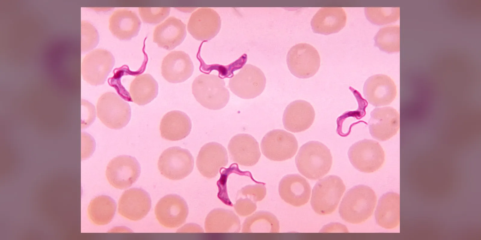
Human African trypanosomiasis, commonly called sleeping sickness, is an infection caused by microscopic parasites called trypanosomes. It is spread by the bite of infected tsetse flies, found only in rural areas of sub-Saharan Africa.
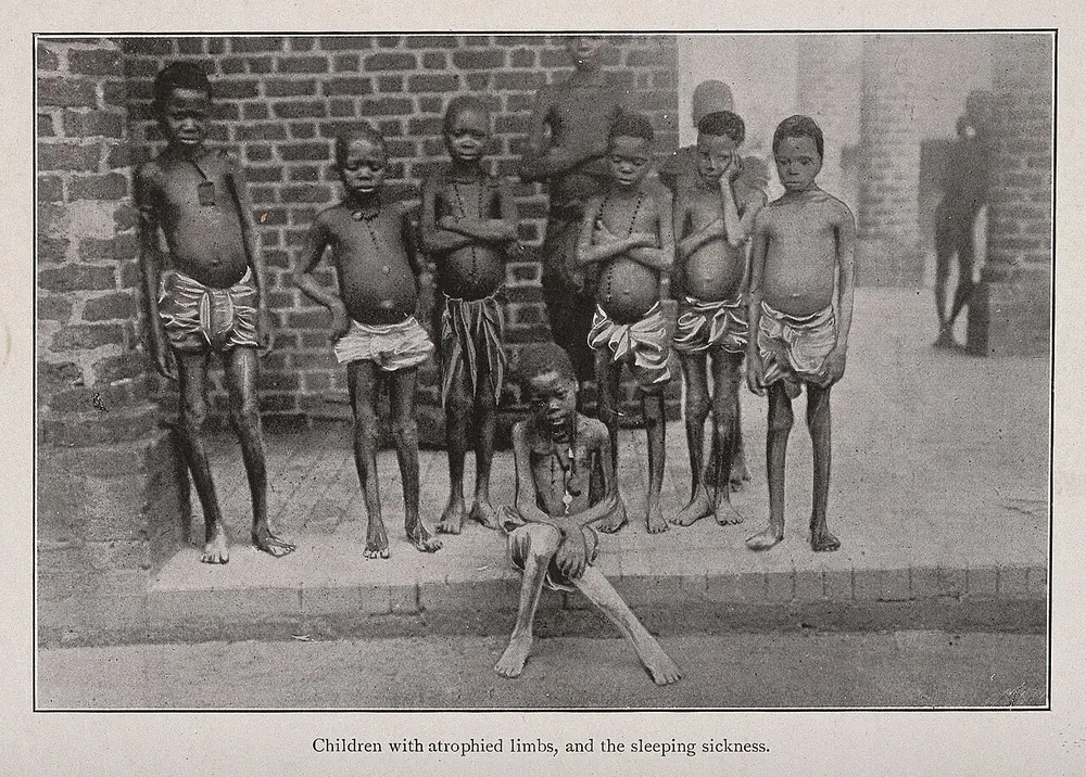
The disease has two stages. In the first stage, the parasites multiply in the blood and lymph glands, causing fever, headaches, joint pains and swollen glands. In the second stage, they cross into the brain and spinal cord, causing confusion, changes in behaviour, poor coordination and the characteristic disturbance of the sleep cycle that gives the disease its name. Without treatment, the disease is almost always fatal.
Thanks to sustained control programmes, case numbers have fallen dramatically, and the disease has been targeted for elimination. However, it still affects remote communities, and travellers to game parks and rural areas in endemic countries can occasionally be infected. Newer, simpler treatments, including oral medicines, have transformed care.
When to get urgent help
Seek urgent medical care if you develop:
- Fever, headache or a painful sore after a trip to rural sub-Saharan Africa
- Confusion, unusual sleepiness, behaviour changes or seizures in someone who has lived in or visited an endemic area
Causes and risk factors
Sleeping sickness is caused by two subspecies of the parasite Trypanosoma brucei:
- T. b. gambiense: found in West and Central Africa; causes more than 90 percent of cases; produces a slowly progressive, chronic illness; humans are the main reservoir
- T. b. rhodesiense: found in East and Southern Africa; causes an acute, rapidly progressive illness; mainly carried by wild and domestic animals, such as antelope and cattle
The parasites are transmitted by the bite of infected tsetse flies (Glossina species), which live in vegetation near rivers, lakes, forests and savannah. Other, rarer routes include mother-to-child transmission during pregnancy, blood transfusion, organ transplantation and laboratory accidents.
People at higher risk include those who farm, fish, hunt, herd livestock or collect water and wood in endemic areas, as well as visitors to national parks and game reserves.
How it develops (pathophysiology)
The parasite and its life cycle
Trypanosomes are single-celled parasites with a long, slender body and a whip-like flagellum that allows them to swim through blood and tissue fluid. They live outside cells (extracellularly), floating in the blood, lymph and, eventually, the fluid around the brain.
The life cycle involves both the tsetse fly and a mammal:
- When an infected tsetse fly bites, it injects metacyclic trypomastigotes from its salivary glands into the skin.
- In the skin, the parasites multiply and transform into bloodstream trypomastigotes.
- They spread through the lymphatic system and enter the bloodstream, where they multiply by dividing in two.
- Some parasites change into a non-dividing "stumpy" form that is adapted to survive in the fly.
- When a tsetse fly bites an infected person or animal, it takes up these parasites, which develop in the fly's gut and migrate to its salivary glands over several weeks, ready to infect a new host.
How the parasite evades the immune system
The surface of each trypanosome is covered by a dense coat made of about ten million copies of a single protein called the variant surface glycoprotein (VSG). The immune system recognises this coat and produces antibodies that destroy most parasites. However, the trypanosome genome contains more than a thousand different VSG genes. A small number of parasites in each generation switch to expressing a new VSG, a process called antigenic variation. These parasites are not recognised by existing antibodies and multiply, producing a new wave of infection. The immune system responds again, and the cycle repeats.
This explains the waves of fever and parasite numbers in the blood. Repeated stimulation also drives very high levels of a type of antibody called IgM, which is a useful diagnostic clue, and contributes to immune exhaustion over time.
Humans have some natural protection: a component of high-density lipoprotein in human blood called trypanosome lytic factor, containing a protein called apolipoprotein L1 (APOL1), kills many animal trypanosomes. T. b. rhodesiense produces a protein called serum resistance-associated protein (SRA) that neutralises APOL1, and T. b. gambiense uses other mechanisms, allowing them to infect humans.
How the disease develops
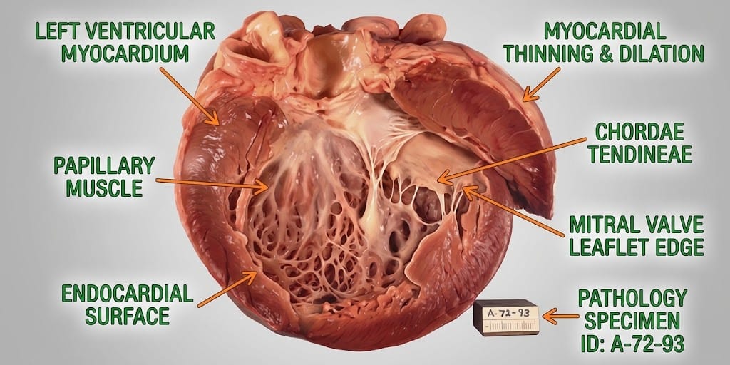
Stage 1: haemolymphatic stage. After the bite, a painful, red, swollen nodule called a trypanosomal chancre may develop at the bite site within one to two weeks, especially with rhodesiense. Parasites then spread to the lymph nodes and blood. The immune system releases inflammatory cytokines, such as tumour necrosis factor and interferon gamma, causing fever, headache, joint pains and itching. Lymph nodes enlarge as immune cells multiply in response. Inflammation can affect the heart (myocarditis), liver, spleen, kidneys and endocrine glands.
Stage 2: meningoencephalitic stage. Over weeks (rhodesiense) or months to years (gambiense), parasites cross the blood-brain barrier, probably by passing between the cells lining small blood vessels in areas where the barrier is weaker, such as the choroid plexus and around brain vessels. Once in the central nervous system, they trigger:
- Inflammation of the meninges and brain tissue (meningoencephalitis), with cuffs of immune cells around blood vessels (perivascular cuffing)
- Activation of brain support cells, astrocytes and microglia, which release cytokines and other substances
- Appearance of large plasma cells filled with antibodies, called Mott cells
- Damage to the white matter and to nerve cells, especially in deep brain areas
Why the sleep disturbance happens
The brain's sleep-wake cycle is controlled by the hypothalamus, especially the suprachiasmatic nucleus, which acts as the body's master clock, along with other hypothalamic and brainstem centres. In sleeping sickness, inflammation in and around the hypothalamus disrupts these circuits. Inflammatory cytokines and molecules released by the parasites, such as prostaglandin D2, which promotes sleep, also affect the sleep centres.
The result is not simply excessive sleepiness, but a fragmentation and reversal of the sleep-wake cycle: people sleep in short episodes throughout the day and night, often experiencing daytime sleepiness and night-time insomnia. Hormone rhythms such as cortisol and prolactin are also disrupted.
Why the other symptoms happen
- Fever, headache, joint and muscle pains reflect cytokine release during waves of parasites in the blood.
- Swollen lymph nodes, particularly at the back of the neck (Winterbottom's sign, classic in gambiense infection), result from immune activation in the lymph nodes.
- Itching and rashes are caused by immune reactions in the skin.
- Anaemia and weight loss are due to chronic inflammation, destruction of red blood cells and poor nutrition.
- Confusion, irritability, personality changes and psychiatric symptoms reflect inflammation in the brain, particularly in the frontal lobes and deep structures.
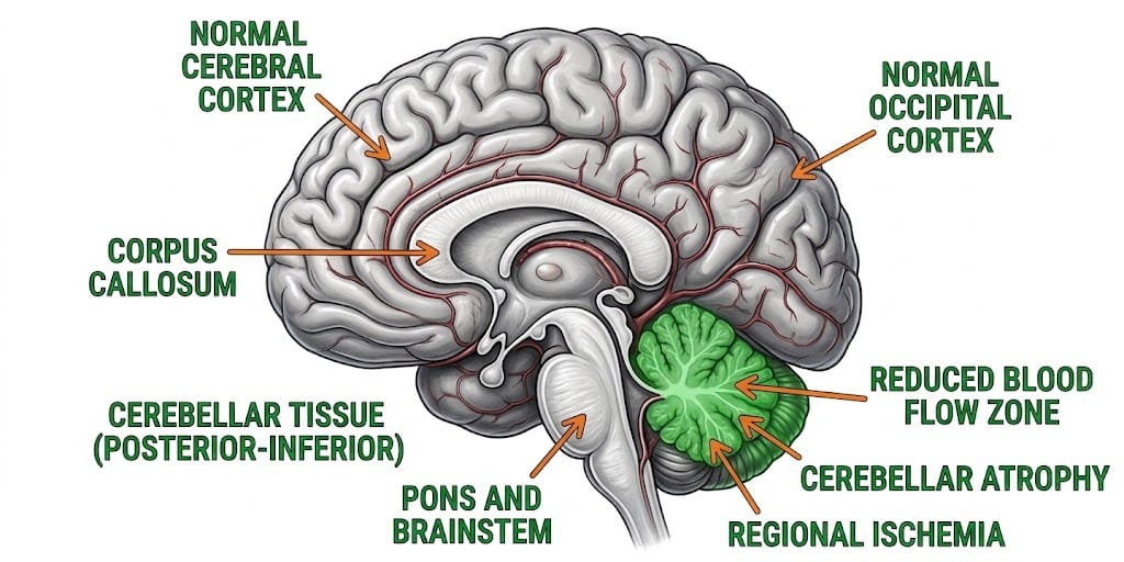
- Tremor, muscle stiffness, poor coordination and abnormal movements arise from damage to deep brain areas such as the basal ganglia and to connections with the cerebellum.
- Exaggerated pain response (Kerandel's sign), where light pressure causes severe pain, reflects nerve involvement.
- Heart symptoms, such as palpitations or breathlessness, occur with myocarditis, especially in rhodesiense.
How complications develop
Without treatment, progressive brain inflammation leads to seizures, coma and death. Malnutrition, secondary infections and heart failure contribute to death in many cases. Even after treatment, some people have lasting neurological or psychiatric problems. Some older treatments, particularly melarsoprol, can cause a severe reaction called post-treatment reactive encephalopathy, a sudden worsening of brain inflammation that can be fatal.
How treatment acts on the mechanism
- Fexinidazole, an oral medicine, is converted into active compounds that damage parasite DNA and proteins; it crosses into the brain, allowing treatment of both stages of gambiense infection without hospital injections.
- Acoziborole, a newer single-dose oral treatment for gambiense infection, inhibits an enzyme needed for the parasite's RNA processing.
- Pentamidine treats first-stage gambiense infection by interfering with parasite DNA and metabolism; it does not cross well into the brain.
- Suramin treats first-stage rhodesiense infection by inhibiting parasite enzymes.
- Nifurtimox-eflornithine combination therapy (NECT): eflornithine blocks an enzyme called ornithine decarboxylase that parasites need to make polyamines for growth, while nifurtimox generates toxic molecules inside the parasite; used for second-stage gambiense disease.
- Melarsoprol, an arsenic compound that crosses the blood-brain barrier, is now used mainly for second-stage rhodesiense disease where alternatives are not available.
- Fexinidazole is increasingly used for rhodesiense infection as well, reducing reliance on melarsoprol.
Types
- Gambiense HAT (T. b. gambiense): chronic, progressing over months to years; West and Central Africa
- Rhodesiense HAT (T. b. rhodesiense): acute, progressing over weeks; East and Southern Africa
Each type has:
- Stage 1 (haemolymphatic): parasites in blood and lymph
- Stage 2 (meningoencephalitic): parasites in the central nervous system
Signs and symptoms
Stage 1:
- A painful, red swelling at the bite site (chancre), more common with rhodesiense
- Intermittent fever
- Headache
- Joint and muscle pains
- Swollen lymph nodes, especially at the back of the neck
- Itching and rash
- Tiredness and weight loss
Stage 2:
- Disturbed sleep pattern, with daytime sleepiness and night-time insomnia
- Confusion and poor concentration
- Personality changes, irritability, aggression or apathy
- Psychiatric symptoms such as hallucinations
- Tremor, poor coordination and difficulty walking
- Exaggerated pain response
- Seizures and eventually coma
How it is diagnosed
- Screening blood tests, such as the card agglutination test for trypanosomiasis (CATT) or rapid diagnostic tests, for gambiense
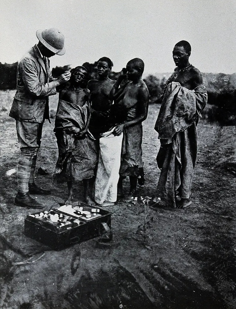
- Microscopy of blood, lymph node fluid or chancre fluid to see parasites; rhodesiense is usually easier to find in blood
- Concentration techniques to increase sensitivity
- Lumbar puncture to examine cerebrospinal fluid for parasites and raised white cells, which determines the stage
- Molecular tests (PCR) in specialist laboratories
- Travel history is crucial for visitors returning from endemic areas
Possible complications
- Brain damage, seizures and coma
- Heart inflammation and heart failure
- Severe weight loss and malnutrition
- Anaemia
- Secondary infections
- Long-term neurological and psychiatric problems
- Death without treatment
- Treatment-related reactions
Goals of treatment
Treatment aims to eliminate the parasites, prevent progression to brain involvement or reverse it, minimise treatment-related harm, and contribute to elimination of the disease through case finding and vector control.
Medicines
- Fexinidazole (oral) for gambiense HAT, both stages in most patients, and increasingly for rhodesiense HAT
- Acoziborole (single oral dose) for gambiense HAT where available
- Pentamidine for stage 1 gambiense HAT
- Suramin for stage 1 rhodesiense HAT
- NECT for severe stage 2 gambiense HAT
- Melarsoprol for stage 2 rhodesiense HAT where newer options are unavailable, with steroids to reduce reaction risk
Treatment choice depends on subspecies, stage, age, pregnancy and local guidelines.
Other treatments
- Hospital care for severely ill patients
- Nutritional support
- Management of seizures and complications
- Active case finding and screening of communities by mobile teams
- Follow-up after treatment to detect relapse
Living with it and prevention
- Wear long-sleeved shirts and trousers in neutral colours (tsetse flies are attracted to bright and dark blue colours)
- Use insect repellent, although it is only partly effective against tsetse flies
- Avoid bushes and vegetation near rivers during the day, when flies are active
- Inspect vehicles for flies before entering
- Support community programmes using traps, insecticide-treated targets and screening
There is no vaccine or preventive medicine.
Monitoring and follow-up
After treatment, people may be followed up with clinical checks and, in some cases, lumbar punctures, to detect relapse. With newer treatments, follow-up schedules have been simplified. Returning travellers with fever should mention their travel history.
Outlook
With early diagnosis and treatment, most people are cured. Second-stage disease has a higher risk of lasting neurological effects. Untreated, the disease is almost always fatal. Elimination efforts have made the disease much rarer.
Special considerations
- Children: may present with developmental delay, seizures or growth failure; newer oral treatments have age and weight restrictions
- Pregnancy: infection can pass to the baby; treatment decisions balance maternal and fetal risks
- Travellers: usually develop rhodesiense infection after visiting game parks; illness can be rapidly severe
- People with HIV may have more complicated disease