
Overview
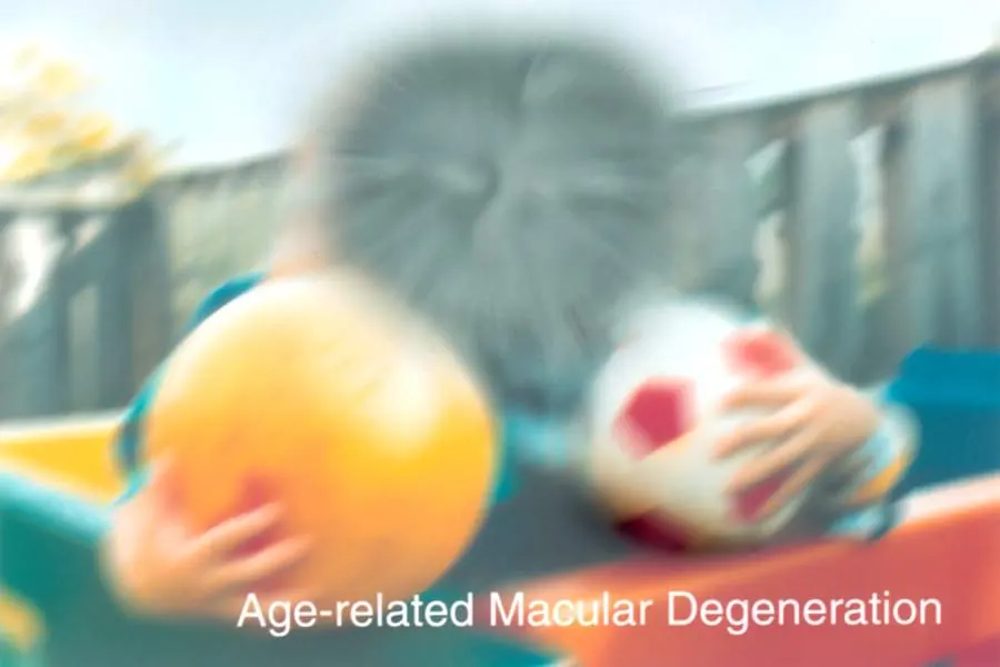
Age-related macular degeneration (AMD) is a common eye condition that affects the macula, the small central part of the retina responsible for sharp, detailed vision. It causes gradual or sometimes sudden loss of central vision, making it difficult to read, recognise faces, drive or see fine detail. Side (peripheral) vision is usually preserved, so AMD does not cause complete blindness.
AMD is the leading cause of severe sight loss in older people in high-income countries. It usually affects people over 50 and becomes more common with age.
There are two main forms. Dry AMD is the most common and progresses slowly over years. Wet AMD develops when abnormal blood vessels grow under the retina and leak, causing rapid vision loss over weeks or months. Wet AMD can be treated effectively with eye injections if caught early, so any sudden change in vision needs urgent attention. Treatments for advanced dry AMD are also emerging.
When to get urgent help
Seek same-day or urgent eye care if you notice:
- Sudden blurring or distortion of central vision
- Straight lines appearing wavy
- A new dark patch in your vision
- Sudden worsening of vision in an eye with AMD
After an eye injection, seek urgent care for increasing eye pain, redness, or reduced vision, which could indicate infection.
Causes and risk factors
AMD results from a combination of ageing, genetics and environment. Risk factors include:
- Age: the strongest risk factor, especially over 60
- Smoking: increases the risk two to four times and speeds progression
- Genetics and family history: variants in genes such as CFH (complement factor H) and ARMS2/HTRA1 greatly influence risk
- Ethnicity: more common in people of European descent
- Cardiovascular disease, high blood pressure and high cholesterol
- Obesity
- Diet low in leafy green vegetables, fruits and oily fish
- Female sex, possibly because women live longer
- Light exposure may contribute, though evidence is less clear
How it develops (pathophysiology)
Normal structure and function of the macula
The retina is the light-sensitive layer at the back of the eye. At its centre is the macula, about 5 to 6 mm across, and in the middle of the macula is the fovea, a small pit packed with cone photoreceptors, which provide colour vision and fine detail. Elsewhere, rod photoreceptors dominate, providing vision in dim light and peripheral vision.
Photoreceptors sit on the retinal pigment epithelium (RPE), a single layer of cells with vital support roles:
- Recycling visual pigment: the RPE regenerates the light-sensitive molecule (retinal) needed for photoreceptors to keep working
- Phagocytosis: every day, photoreceptors shed the tips of their outer segments, which contain damaged membranes and proteins; the RPE engulfs and digests them
- Transport: the RPE carries nutrients and oxygen from the blood to photoreceptors and removes waste and fluid
- Barrier: tight junctions between RPE cells form the outer blood-retinal barrier
- Light absorption: melanin in the RPE absorbs stray light and protects against oxidative damage
Beneath the RPE lies Bruch's membrane, a thin, multi-layered sheet that acts as a filter between the RPE and the choroid, a dense network of blood vessels (the choriocapillaris) that supplies the outer retina. The macula has one of the highest metabolic rates in the body and is exposed to intense light and high oxygen levels, which makes it particularly vulnerable to oxidative stress.
The macula also contains yellow pigments, lutein and zeaxanthin, obtained from the diet, which filter blue light and act as antioxidants.
What goes wrong
AMD develops through several interacting processes:
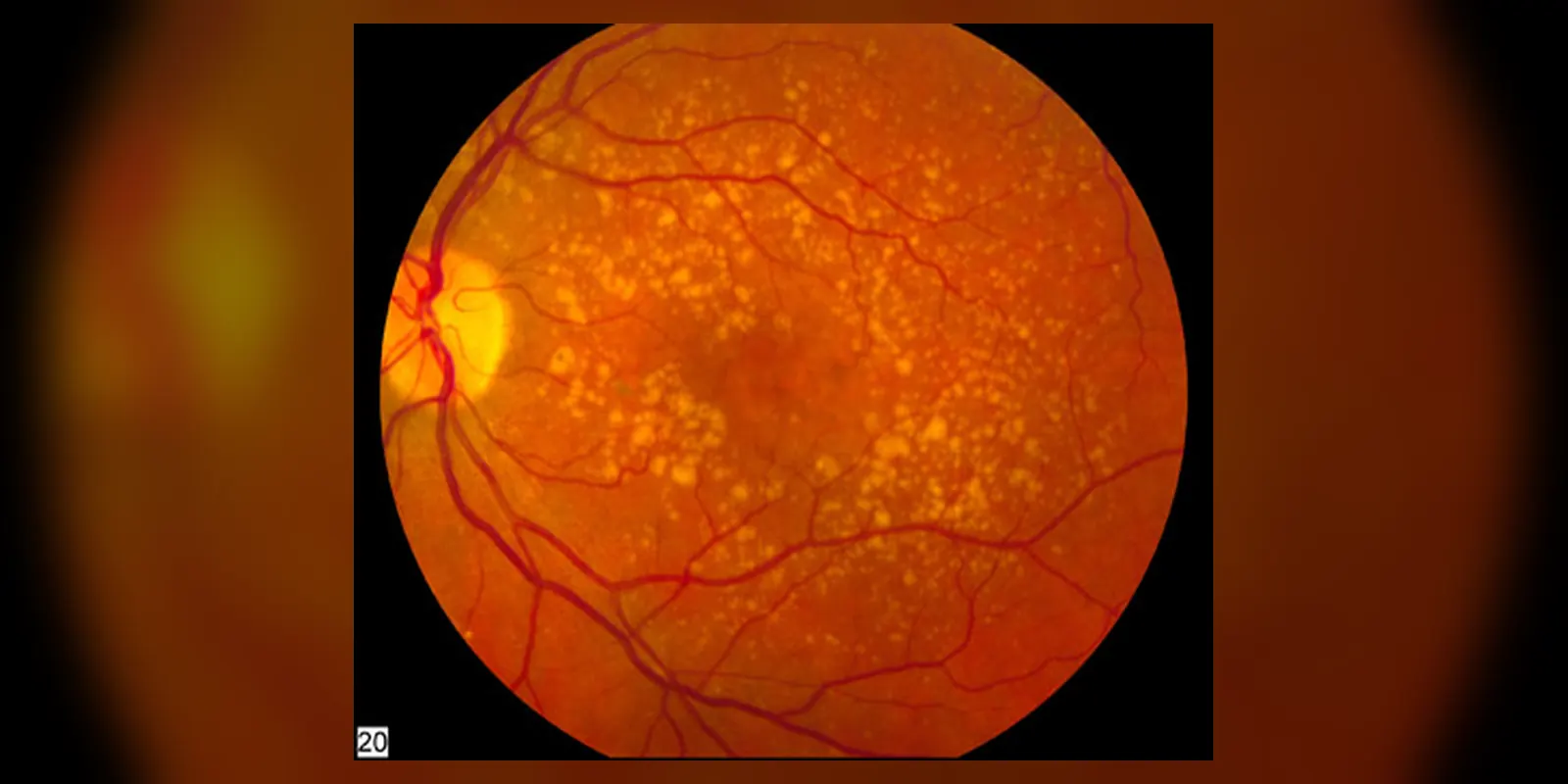
1. Accumulation of waste and drusen. With age, the RPE becomes less efficient at digesting shed photoreceptor material. A fluorescent waste pigment called lipofuscin accumulates in RPE cells. One component, A2E, is toxic when exposed to light, generating reactive oxygen species and impairing cellular function. Waste products, lipids, proteins and inflammatory molecules accumulate between the RPE and Bruch's membrane, forming yellow deposits called drusen. Bruch's membrane thickens and becomes less permeable, impairing the exchange of nutrients and waste.
2. Oxidative stress. High oxygen levels, intense light exposure and high metabolic activity generate reactive oxygen species. Smoking adds more oxidants and reduces antioxidant levels. Over time, damage to DNA, proteins and lipids in the RPE and photoreceptors builds up, and mitochondria become dysfunctional.
3. Complement and inflammation. The complement system, part of the innate immune system, normally clears debris and microbes but must be carefully regulated. Variants in genes such as CFH, which encodes a key complement inhibitor, lead to excessive complement activation. Complement proteins are found in drusen and contribute to chronic low-grade inflammation, damaging the RPE and choriocapillaris. Immune cells, including macrophages and microglia, are drawn to the area.
4. Choroidal blood flow decline. The choriocapillaris thins with age and in AMD, reducing oxygen and nutrient supply to the RPE and photoreceptors.
How dry AMD progresses
In early and intermediate AMD, drusen increase in size and number, and pigment changes appear in the RPE. Vision is often normal or mildly affected. In late dry AMD, called geographic atrophy, patches of RPE cells die. Without RPE support, the overlying photoreceptors and underlying choriocapillaris also die. These atrophic areas enlarge slowly, often first sparing the fovea and then spreading into it, leading to gradual central vision loss.
How wet AMD develops
In neovascular (wet) AMD, the damaged, oxygen-starved RPE and surrounding tissue release vascular endothelial growth factor (VEGF), a signalling protein that stimulates new blood vessel growth. Inflammation and breaks in Bruch's membrane allow abnormal vessels from the choroid to grow into the space under the RPE or retina, a process called choroidal neovascularisation (CNV). These new vessels are fragile and leaky:
- Fluid leaks into and under the retina, causing swelling and distortion
- Blood can bleed under the retina
- Fibrous scarring eventually forms, called a disciform scar, permanently destroying the macula
A related form, polypoidal choroidal vasculopathy, and retinal angiomatous proliferation involve abnormal vessels in slightly different locations.
Why the symptoms happen
- Blurred central vision results from photoreceptor dysfunction and loss in the macula.
- Distorted vision (metamorphopsia), where straight lines look wavy or bent, occurs in wet AMD when fluid lifts and distorts the retina, so photoreceptors are displaced from their normal positions.
- A dark or empty patch in the centre of vision (central scotoma) reflects areas of photoreceptor death or scarring.
- Difficulty reading and recognising faces reflects loss of the fovea's fine detail.
- Needing brighter light and slow adaptation to darkness occur because RPE dysfunction slows the regeneration of visual pigment, especially in rod photoreceptors near the macula.
- Reduced colour and contrast sensitivity reflect cone damage.
- Visual hallucinations (Charles Bonnet syndrome), such as seeing patterns or people that are not there, occur in some people when the brain's visual areas receive reduced input and generate their own images. They are not a sign of mental illness.
How complications develop
Untreated wet AMD can lead to rapid, severe loss of central vision within months as bleeding and scarring destroy the macula. Geographic atrophy progresses more slowly but relentlessly. Reduced vision leads to difficulty reading, driving and recognising faces, falls, depression, loss of independence and social isolation. If one eye has late AMD, the other eye has a higher risk of developing it.
How treatment acts on the mechanism
- Anti-VEGF injections (such as ranibizumab, aflibercept, bevacizumab and faricimab) block VEGF, stopping abnormal vessel growth and leakage. Fluid resolves, the retina flattens, and vision often stabilises or improves. Faricimab also blocks angiopoietin-2, which further stabilises vessels.
- Complement inhibitors (such as pegcetacoplan and avacincaptad pegol), approved in some countries for geographic atrophy, block complement components C3 or C5, slowing the growth of atrophic areas, though they do not restore vision.
- AREDS2-formula supplements containing vitamins C and E, zinc, copper, lutein and zeaxanthin provide antioxidant support and reduce the risk of progression from intermediate to late AMD.
- Stopping smoking reduces oxidative stress and improves choroidal blood flow.
- Photodynamic therapy, used in some cases, closes abnormal vessels using a light-activated drug.
- Low vision aids make the best use of remaining peripheral vision.
Types
- Early AMD: medium-sized drusen, usually no symptoms
- Intermediate AMD: large drusen and/or pigment changes; may have mild symptoms
- Late AMD:
- Dry (atrophic) AMD / geographic atrophy: slow loss of RPE and photoreceptors
- Wet (neovascular) AMD: abnormal blood vessel growth with leakage and bleeding; about 10 to 15 percent of AMD cases but responsible for much severe sight loss
Signs and symptoms
- Blurred or reduced central vision
- Straight lines appearing wavy or distorted
- A dark, blurry or empty area in the centre of vision
- Difficulty reading, even with glasses
- Difficulty recognising faces
- Needing brighter light for close work
- Colours seeming less vivid
- Slow adjustment to dim light
- Seeing things that are not there (Charles Bonnet syndrome)
Wet AMD often causes sudden distortion or loss of vision over days to weeks.
How it is diagnosed
- Visual acuity test using a letter chart
- Amsler grid: a grid of lines to detect distortion or missing areas
- Dilated eye examination to look for drusen, pigment changes, atrophy, fluid or bleeding
- Optical coherence tomography (OCT): a quick, non-invasive scan showing retinal layers, drusen, fluid and atrophy
- OCT angiography: shows abnormal blood vessels without dye
- Fluorescein or indocyanine green angiography: dye injected into a vein to show leaking vessels
- Fundus autofluorescence imaging to map geographic atrophy
Possible complications
- Severe loss of central vision
- Difficulty reading, driving and recognising faces
- Falls and injuries
- Depression and anxiety
- Social isolation and loss of independence
- Charles Bonnet syndrome
- Side effects of injections, such as eye infection (endophthalmitis), raised eye pressure or retinal detachment (rare)
Goals of treatment
The goals are to detect and treat wet AMD quickly to preserve vision, slow the progression of dry AMD, maximise use of remaining vision, and support independence and wellbeing.
Medicines
- Anti-VEGF injections into the eye for wet AMD, given regularly, often monthly at first, then at intervals tailored to response ("treat and extend")
- Complement inhibitor injections for geographic atrophy, where approved
- AREDS2 supplements for intermediate AMD or late AMD in one eye, on the advice of an eye specialist
- Photodynamic therapy in selected cases
Other treatments
- Low vision rehabilitation: magnifiers, bright task lighting, large-print materials, screen readers and electronic magnifiers
- Eccentric viewing training to use healthy parts of the retina
- Occupational therapy and home adaptations to reduce falls
- Registration as sight-impaired to access support services
- Counselling and support groups
Living with it and prevention
- Stop smoking
- Eat a diet rich in leafy green vegetables (such as spinach and kale), fruit, nuts and oily fish
- Keep blood pressure and cholesterol under control
- Stay physically active and maintain a healthy weight
- Wear sunglasses in bright light
- Have regular eye tests, especially if you are over 60 or have a family history
- Check each eye separately with an Amsler grid regularly if you have AMD
Monitoring and follow-up
People with intermediate AMD should have regular eye checks and self-monitor at home. People receiving anti-VEGF injections need regular OCT scans to guide treatment intervals. The other eye is also monitored for signs of wet AMD.
Outlook
Dry AMD usually progresses slowly, and many people keep useful vision for years. Wet AMD treated promptly with anti-VEGF injections can often be stabilised, and some people gain vision. Without treatment, wet AMD often leads to severe central vision loss. AMD does not cause total blindness because peripheral vision is preserved.
Special considerations
- Drivers: AMD can affect legal fitness to drive; vision standards must be checked
- People with dementia: may find frequent injections challenging and need extra support
- People living alone: low vision and falls prevention services are especially important