
Overview
Alpha-1 antitrypsin deficiency (AATD) is an inherited condition in which the body does not make enough of a protein called alpha-1 antitrypsin (AAT), or makes an abnormal form of it. AAT is made in the liver and travels to the lungs, where it protects the delicate air sacs from being damaged by enzymes released by white blood cells.
Without enough AAT, the lungs are gradually damaged, causing emphysema and COPD, often at a younger age than usual, typically between 30 and 50, and especially in people who smoke. In some forms, the abnormal protein gets stuck in liver cells, causing liver disease in babies, children or adults. AATD is under-recognised; many people are diagnosed with asthma or COPD for years before the cause is found. Stopping smoking, protecting the lungs and liver, and in some cases protein replacement therapy can slow progression.
When to get urgent help
Seek urgent help for severe breathlessness, blue lips, coughing up blood, confusion, vomiting blood, black stools, or worsening jaundice.
Causes and risk factors
AATD is caused by changes (variants) in the SERPINA1 gene, inherited from both parents (co-dominant inheritance). The common gene versions are:
- M: normal
- Z: the most common severe variant; the protein misfolds and builds up in the liver
- S: a milder variant
People with ZZ have severe deficiency. SZ carries moderate risk, especially in smokers. MZ carriers have slightly reduced levels and increased risk if they smoke. Rare null variants produce no protein. The Z variant is most common in people of Northern European descent.
Factors that increase lung damage include smoking, dust, fumes and repeated chest infections. Alcohol and obesity add to liver risk.
How it develops (pathophysiology)
Normal role of alpha-1 antitrypsin
When the lungs face infection or irritants, neutrophils (white blood cells) move into the air sacs and release an enzyme called neutrophil elastase. Elastase breaks down bacteria and debris but can also digest elastin, the stretchy protein that lets the air sacs expand and recoil. Alpha-1 antitrypsin, a member of the serpin family of protease inhibitors, is made mainly by liver cells, released into the blood and diffuses into the lungs, where it binds to elastase and switches it off. This keeps a balance between protein breakdown and protection.
What goes wrong in the lungs
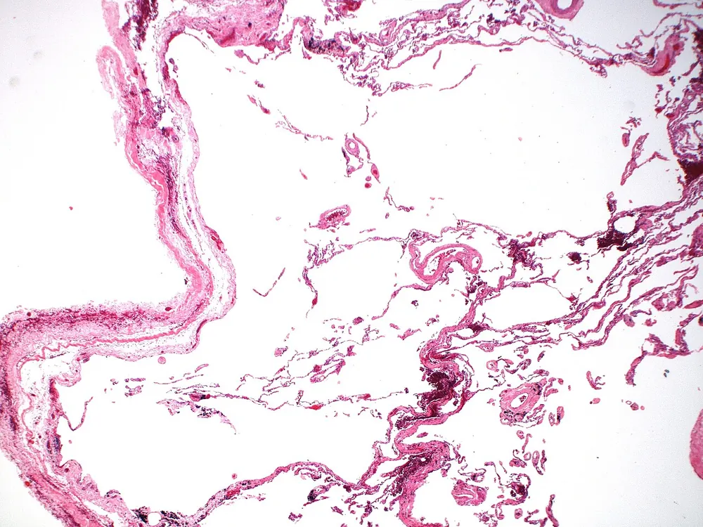
With low AAT levels (a protease-antiprotease imbalance), elastase acts unchecked. Over years, it digests the walls of the alveoli. Small air sacs merge into larger, floppy spaces, a process called emphysema. In AATD, damage typically affects the lower parts of the lungs first (panlobular emphysema), unlike smoking-related emphysema, which usually starts at the top. Smoking makes this far worse: it attracts more neutrophils and oxidises the remaining AAT so that it stops working. The Z protein also forms clumps (polymers) that may themselves attract neutrophils, adding to inflammation.
What goes wrong in the liver
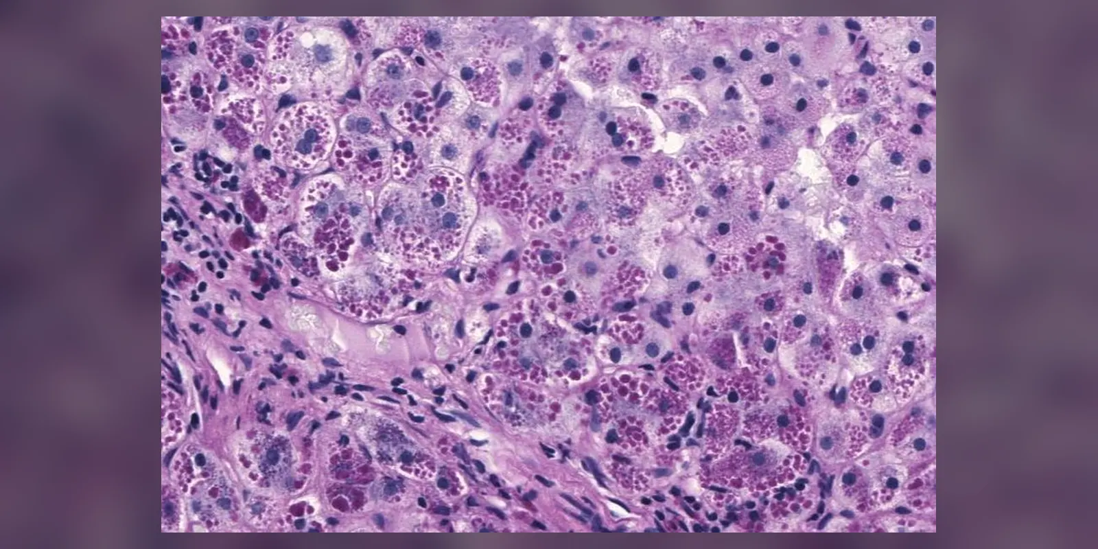
The Z variant makes AAT fold incorrectly. Instead of being released into the blood, many Z molecules link together into chains (polymers) inside the endoplasmic reticulum of liver cells. These accumulate as globules that stress and damage the cells. The liver tries to clear them through autophagy, but over time inflammation, fibrosis and cirrhosis can develop. Liver cell damage also raises the risk of liver cancer. Null variants cause lung disease but not liver disease, because no protein is made to accumulate.
Why the symptoms happen
- Breathlessness on exertion results from loss of air sacs and stiffening of airways, so less oxygen is exchanged and air is trapped.
- Wheeze and cough reflect airway inflammation and narrowing.
- Frequent chest infections occur because damaged airways clear bacteria poorly.
- Jaundice in newborns and liver problems result from protein build-up in liver cells.
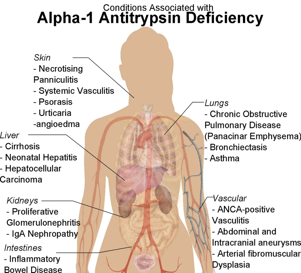
- Panniculitis, a rare painful skin condition, results from unchecked enzyme activity in fat tissue.
How treatment acts on the mechanism
Stopping smoking removes the strongest driver of lung damage. AAT augmentation therapy, given as weekly infusions of purified AAT, raises levels in the blood and lungs, slowing the loss of lung tissue in selected people. Vaccinations and prompt treatment of infections reduce neutrophil surges. Liver disease is managed by avoiding alcohol and, in severe cases, transplantation, which cures the deficiency because the new liver makes normal AAT.
Types
- Lung-predominant disease: emphysema and COPD in adults
- Liver-predominant disease: neonatal jaundice and hepatitis, childhood liver disease, or cirrhosis in adults
- Mixed disease
- Classified by genotype (ZZ, SZ, MZ, null)
Signs and symptoms
- Shortness of breath, especially on exertion
- Wheezing
- Long-lasting cough with or without sputum
- Frequent chest infections
- Reduced exercise tolerance and tiredness
- Asthma that does not respond well to treatment
- Yellowing of the skin and eyes (jaundice), especially in newborns
- Swollen abdomen, easy bruising or vomiting blood in liver disease
- Rarely, painful red lumps under the skin (panniculitis)
How it is diagnosed
- Blood test for AAT level
- Genetic testing (genotyping or phenotyping) to identify variants
- Lung function tests (spirometry)
- Chest CT scan to show emphysema, typically in the lower lungs
- Liver tests, ultrasound and elastography to assess liver damage
- Testing of family members once a diagnosis is made
Guidelines recommend testing everyone with COPD, adult-onset asthma that is hard to treat, unexplained liver disease, or panniculitis.
Possible complications
- Emphysema and COPD
- Bronchiectasis
- Respiratory failure
- Cirrhosis and liver failure
- Liver cancer
- Panniculitis
- Vasculitis, rarely
Goals of treatment
The goals are to stop or slow lung and liver damage, relieve symptoms, prevent infections and support quality of life.
Medicines
- Inhalers: bronchodilators and sometimes inhaled steroids, as for COPD
- AAT augmentation therapy (intravenous AAT) for selected people with severe deficiency and lung disease, where available
- Antibiotics for chest infections
- Vaccinations against influenza, pneumococcus and COVID-19
- Treatment of liver complications
Other treatments
- Stopping smoking, the single most important measure
- Pulmonary rehabilitation
- Oxygen therapy if oxygen levels are low
- Lung volume reduction or lung transplantation in advanced disease
- Liver transplantation in end-stage liver disease
- Genetic counselling for families
Living with it and prevention
- Never smoke and avoid second-hand smoke
- Avoid dust, fumes and pollution at work and home
- Limit or avoid alcohol
- Keep a healthy weight
- Stay active and exercise regularly
- Get vaccinated
- Treat chest infections early
- Encourage family members to be tested
Monitoring and follow-up
Regular lung function tests, liver tests and liver scans are recommended. People with liver involvement may need surveillance for liver cancer. Children with liver disease need specialist follow-up.
Outlook
Outlook varies widely. People with severe deficiency who never smoke may have near-normal life expectancy, while smoking greatly increases the risk of early severe lung disease. Liver disease affects a minority.
Special considerations
- Newborns and children: may present with prolonged jaundice or liver problems
- Smokers with MZ or SZ genotypes: higher risk of COPD
- Family members: testing allows early prevention