
Overview
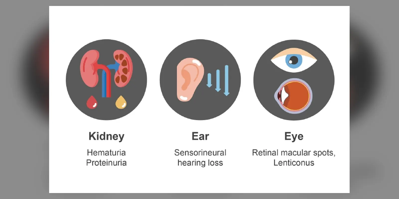
Alport syndrome is an inherited condition caused by changes in the genes for a type of collagen called type IV collagen. This collagen forms part of the thin filtering membranes in the kidneys, the inner ear and the eyes. When it is abnormal, these structures gradually fail.
The main feature is progressive kidney disease, which usually starts with blood in the urine in childhood and can lead to kidney failure, often in early adulthood in men with the most common form. Many people also develop hearing loss and eye changes. Alport syndrome cannot be cured, but early treatment with medicines that protect the kidneys can significantly delay kidney failure, and kidney transplantation works well when needed.
When to get urgent help
Seek urgent help for very little urine, severe swelling, breathlessness, severe headache with very high blood pressure, or signs of transplant rejection such as fever and pain over the transplant.
Causes and risk factors
Alport syndrome is caused by changes in genes that make the type IV collagen alpha-3, alpha-4 and alpha-5 chains:
- X-linked Alport syndrome (COL4A5 gene): about 80 percent of cases; males are usually more severely affected, while females have variable disease
- Autosomal recessive Alport syndrome (COL4A3 or COL4A4 genes): both copies are affected; severe in males and females
- Autosomal dominant Alport syndrome: one changed copy of COL4A3 or COL4A4; usually milder and later onset
A family history of kidney disease, blood in urine or hearing loss is common, but some cases arise from new gene changes.
How it develops (pathophysiology)
Normal structure of the kidney filter
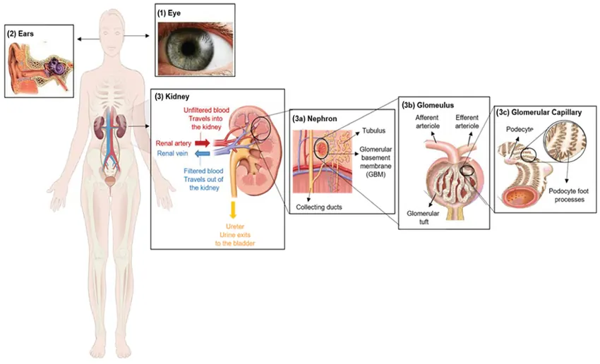
Each kidney contains about a million filters called glomeruli. Blood is filtered through three layers: the cells lining the capillaries, the glomerular basement membrane (GBM), and specialised cells called podocytes. The GBM is a fine mesh made mainly of type IV collagen. In healthy adults, the GBM contains a strong network made of alpha-3, alpha-4 and alpha-5 chains twisted together. This network is resistant to stress and keeps blood cells and protein in the blood while allowing water and waste through.
Similar membranes, also rich in the same collagen network, are found in the cochlea of the inner ear and in the lens capsule and retina of the eye.
What goes wrong
In Alport syndrome, a gene change prevents normal alpha-3/alpha-4/alpha-5 collagen from being made or assembled. The body continues to use the immature alpha-1/alpha-2 collagen network, which is weaker and more easily damaged. Over time:
- The GBM becomes thin, then irregularly thickened and split, with a characteristic "basket-weave" appearance under the electron microscope.
- Weak areas allow red blood cells to leak into the urine, causing haematuria from early childhood.
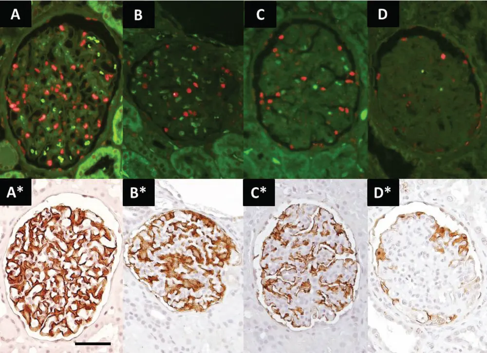
- Mechanical stress damages podocytes, which detach, so protein leaks into the urine (proteinuria).
- Damaged glomeruli release inflammatory and scarring signals such as transforming growth factor beta, leading to glomerulosclerosis and scarring of the tissue between tubules (interstitial fibrosis).
- As more glomeruli fail, kidney function declines towards chronic kidney disease and kidney failure.
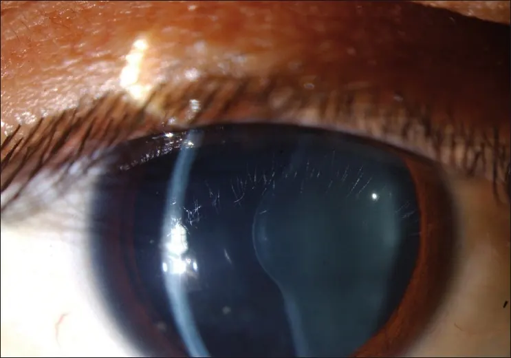
In the inner ear, faulty basement membranes in the cochlea affect how sound vibrations are converted into nerve signals, causing sensorineural hearing loss, usually affecting high frequencies first, and typically appearing in late childhood or adolescence. In the eyes, the thin lens capsule can bulge forward (anterior lenticonus), and flecks appear on the retina.
In X-linked disease, females have one normal and one changed X chromosome. Because one X is randomly switched off in each cell, some areas of their GBM have normal collagen and others do not, which explains their milder and more variable disease.
How treatment acts on the mechanism
ACE inhibitors and angiotensin receptor blockers lower pressure within the glomeruli and reduce protein leakage, slowing scarring and delaying kidney failure, especially when started early. SGLT2 inhibitors may add further protection. Controlling blood pressure reduces stress on the fragile filter. Kidney transplantation replaces failing kidneys; a small number of recipients develop antibodies against the normal collagen in the donor kidney (anti-GBM disease), which is monitored.
Types
- X-linked Alport syndrome
- Autosomal recessive Alport syndrome
- Autosomal dominant Alport syndrome
- Thin basement membrane nephropathy is now often considered part of the Alport spectrum
Signs and symptoms
- Blood in the urine, often invisible but sometimes making urine red or brown, especially after infections
- Protein in the urine, causing foamy urine
- Swelling of the legs and around the eyes
- High blood pressure
- Hearing loss, usually starting in late childhood or the teenage years
- Vision problems from lens or retinal changes
- Symptoms of kidney failure later: tiredness, itching, nausea and poor appetite
How it is diagnosed
- Urine tests for blood and protein
- Blood tests for kidney function
- Genetic testing, now the main way to confirm the diagnosis
- Kidney biopsy with electron microscopy in some cases
- Hearing tests (audiometry)
- Eye examination
- Family testing once a diagnosis is made
Possible complications
- Chronic kidney disease and kidney failure
- High blood pressure
- Hearing loss
- Vision problems
- Anti-GBM disease after kidney transplant, rarely
Goals of treatment
The goals are to slow kidney disease progression, control blood pressure, support hearing and vision, plan for kidney replacement therapy, and support families through genetic counselling.
Medicines
- ACE inhibitors (such as ramipril) or angiotensin receptor blockers, started early when protein appears in the urine
- SGLT2 inhibitors in some people with chronic kidney disease
- Blood pressure control
- Treatment of complications of chronic kidney disease, such as anaemia and bone disease
Other treatments
- Dialysis when kidneys fail
- Kidney transplantation, which usually works well
- Hearing aids
- Eye surgery for lenticonus or cataract if needed
- Genetic counselling for family planning
Living with it and prevention
- Have regular urine and blood pressure checks if Alport syndrome runs in the family
- Keep blood pressure controlled
- Avoid medicines that harm the kidneys, such as regular NSAIDs
- Protect hearing from loud noise
- Eat a healthy diet and stay active
Monitoring and follow-up
Regular monitoring includes urine protein, kidney function, blood pressure, hearing tests and eye checks. Children with Alport syndrome are followed by paediatric kidney specialists.
Outlook
Without treatment, most men with X-linked Alport syndrome reach kidney failure in their twenties or thirties. Early treatment with ACE inhibitors can delay this by many years. Women with X-linked disease and people with autosomal dominant forms usually have milder disease.
Special considerations
- Children: early diagnosis allows treatment to start before significant damage
- Women with X-linked disease: need lifelong monitoring, as kidney disease can develop later
- Pregnancy: kidney function and blood pressure are monitored closely