
Overview
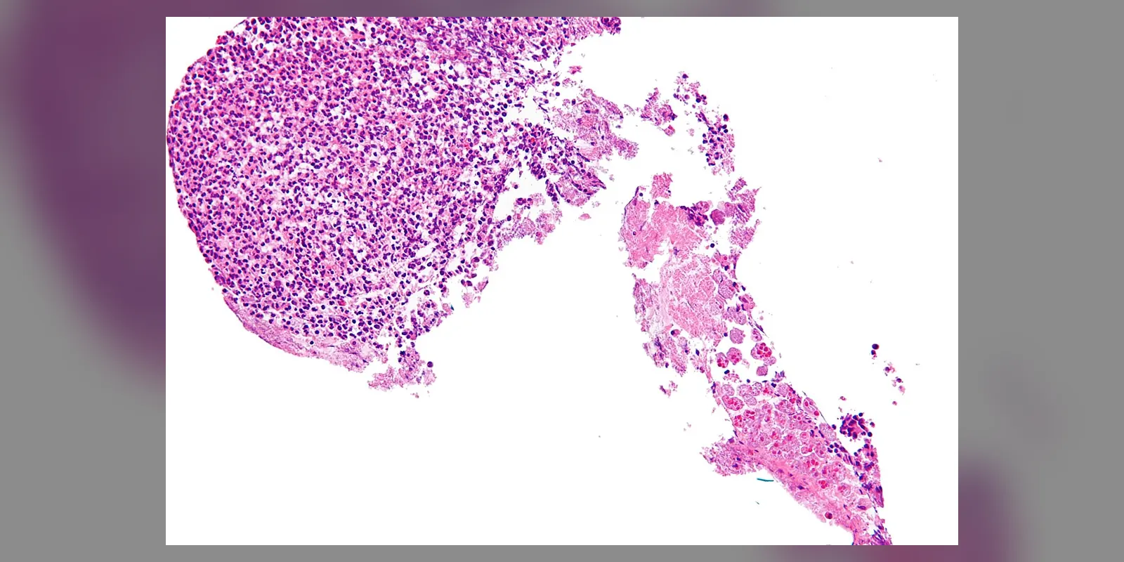
Amoebiasis is an infection caused by the single-celled parasite Entamoeba histolytica. It spreads through food or water contaminated with faeces and is common in areas with poor sanitation, particularly in parts of Asia, Africa and Latin America. Travellers and migrants can also be affected.
Most infected people have no symptoms. In others, the parasite invades the bowel wall, causing amoebic colitis, with diarrhoea that may contain blood and mucus (amoebic dysentery). Occasionally, it spreads through the bloodstream to the liver, forming an amoebic liver abscess. Amoebiasis is treatable with medicines that kill the parasite in the tissues and in the bowel.
When to get urgent help
Seek urgent care for severe abdominal pain, high fever, heavy bloody diarrhoea, signs of dehydration, or pain in the right upper abdomen with fever.
Causes and risk factors
- Swallowing cysts of Entamoeba histolytica in contaminated water, food or on hands
- Poor sanitation and lack of clean water
- Person-to-person spread, including through sexual contact involving the anus
- Travel or migration from endemic areas
- Living in institutions with crowded conditions
Higher risk of severe disease occurs in young children, pregnant women, malnourished people, older adults and those taking steroids.
How it develops (pathophysiology)
Life cycle of the parasite
Entamoeba histolytica exists in two forms:
- Cysts: hardy, infective forms that survive outside the body in water and soil
- Trophozoites: active forms that live and multiply in the large bowel
When cysts are swallowed, they pass through the stomach unharmed and release trophozoites in the small intestine. These move to the colon, where they feed on bacteria and debris. Some form new cysts, which pass in faeces and spread the infection.
What goes wrong
In most people, trophozoites live harmlessly in the bowel. In some, they invade the bowel wall:
- Attachment: trophozoites attach to the colon lining using a surface protein (Gal/GalNAc lectin) that binds to sugars on cells.
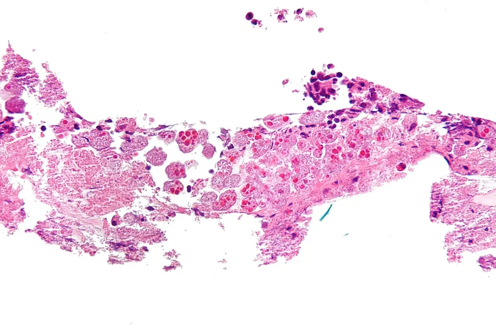
- Killing of cells: they kill human cells by contact, using pore-forming proteins called amoebapores and by triggering cell death. They can then swallow the dead cells and even nibble pieces of living cells (trogocytosis).
- Tissue breakdown: cysteine proteases digest the mucus layer and the matrix holding cells together, allowing deeper invasion.
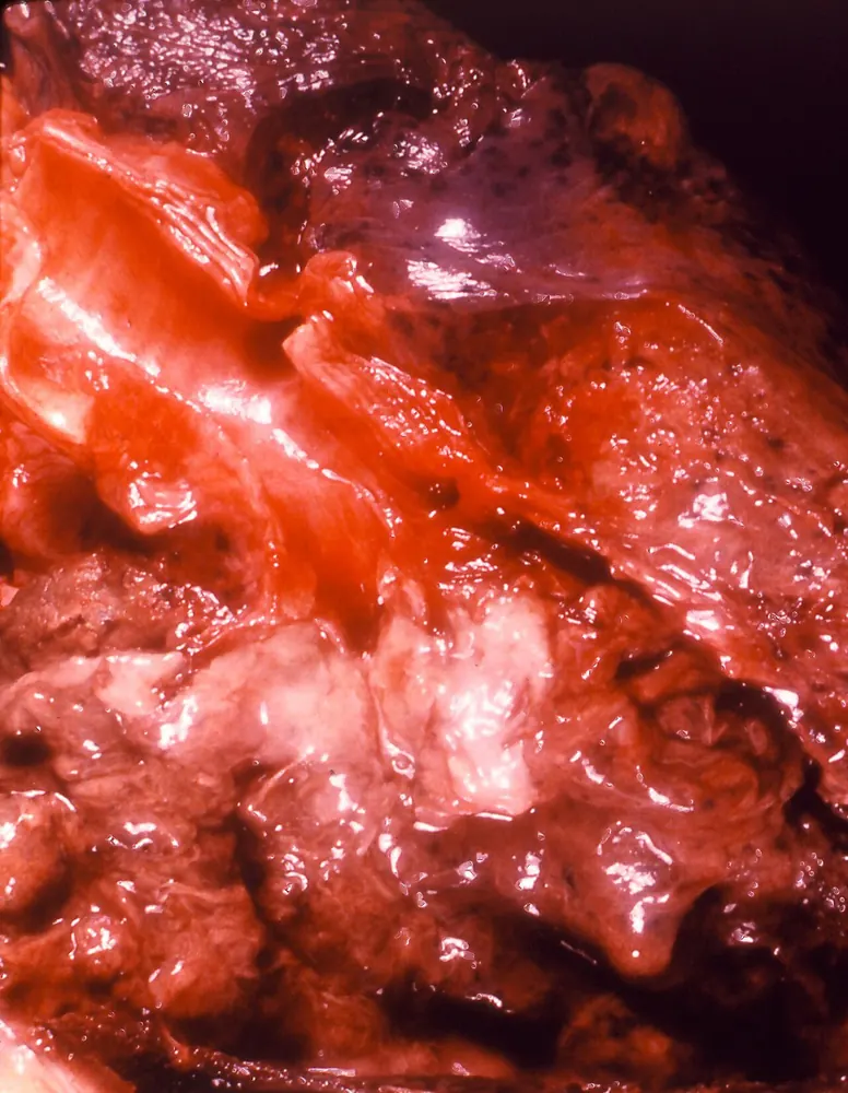
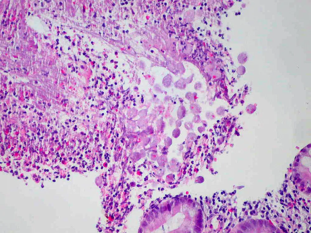
- Ulceration: invasion creates small surface ulcers that widen beneath the surface into flask-shaped ulcers, mainly in the caecum and ascending colon.
- Inflammation: the immune system responds with neutrophils and inflammatory signals, which add to tissue damage.
Trophozoites can enter the portal vein, which drains blood from the gut to the liver. In the liver, they destroy tissue, creating an amoebic liver abscess filled with liquefied dead liver tissue, often described as "anchovy paste". Rarely, infection spreads to the lungs, brain or skin.
Why the symptoms happen
- Diarrhoea results from inflammation and ulceration of the colon lining, impairing water absorption.
- Blood and mucus in stools occur when ulcers bleed and mucus is produced.
- Abdominal pain and cramps come from bowel inflammation.
- Fever and right upper abdominal pain suggest liver abscess, as the liver capsule is stretched.
- Weight loss and tiredness result from chronic infection and inflammation.
How treatment acts on the mechanism
Metronidazole or tinidazole kill trophozoites in the bowel wall and liver. Because they are less effective against parasites inside the bowel, a second medicine, a luminal agent such as paromomycin or diloxanide furoate, is given to clear remaining parasites and cysts, preventing relapse and spread.
Types
- Asymptomatic infection: most common
- Amoebic colitis (amoebic dysentery)
- Amoeboma: a mass in the bowel wall that can mimic cancer
- Amoebic liver abscess
- Other extraintestinal amoebiasis: lung, brain, skin
Signs and symptoms
- Diarrhoea, sometimes with blood and mucus
- Abdominal pain and cramping
- Urgency to open the bowels
- Fever
- Weight loss and tiredness
- Liver abscess: fever, pain in the upper right abdomen or right shoulder, tenderness over the liver, sometimes without diarrhoea
Symptoms often develop gradually over weeks.
How it is diagnosed
- Stool tests: antigen tests or PCR to identify E. histolytica specifically (microscopy cannot reliably distinguish it from harmless species)
- Blood tests for antibodies, especially for liver abscess
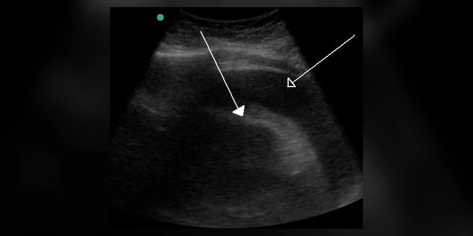
- Liver ultrasound or CT to detect abscesses
- Colonoscopy with biopsy in some cases
- Blood count and inflammatory markers
Possible complications
- Severe colitis with bowel perforation or toxic megacolon
- Amoebic liver abscess, which may rupture into the chest or abdomen
- Dehydration
- Amoeboma causing bowel blockage
- Spread to lungs, heart lining or brain, rarely
Goals of treatment
Treatment aims to kill the parasite in the tissues and bowel, relieve symptoms, prevent complications and stop spread.
Medicines
- Metronidazole or tinidazole for invasive disease
- Followed by a luminal agent (paromomycin or diloxanide furoate)
- Luminal agents alone for asymptomatic carriers
- Fluids for dehydration
Other treatments
- Drainage of liver abscess if large, at risk of rupture, or not improving
- Surgery for bowel perforation or severe complications
- Rehydration
Living with it and prevention
- Drink safe water: boiled, filtered or bottled
- Wash hands with soap after using the toilet and before eating
- Peel fruit and cook vegetables in endemic areas
- Avoid ice and uncooked foods where water may be unsafe
- Improve sanitation in communities
- Treat infected household contacts
Monitoring and follow-up
Stool tests may be repeated after treatment to confirm clearance. Liver abscesses are monitored with ultrasound until they resolve.
Outlook
With treatment, most people recover fully. Liver abscesses usually resolve with medicines alone. Severe complications such as perforation can be life-threatening.
Special considerations
- Children and pregnant women: more prone to severe disease
- People taking steroids: steroids can worsen amoebic colitis, so amoebiasis should be excluded before treating suspected inflammatory bowel disease in people from endemic areas
- Travellers: consider amoebiasis in returning travellers with bloody diarrhoea or liver abscess