
Overview
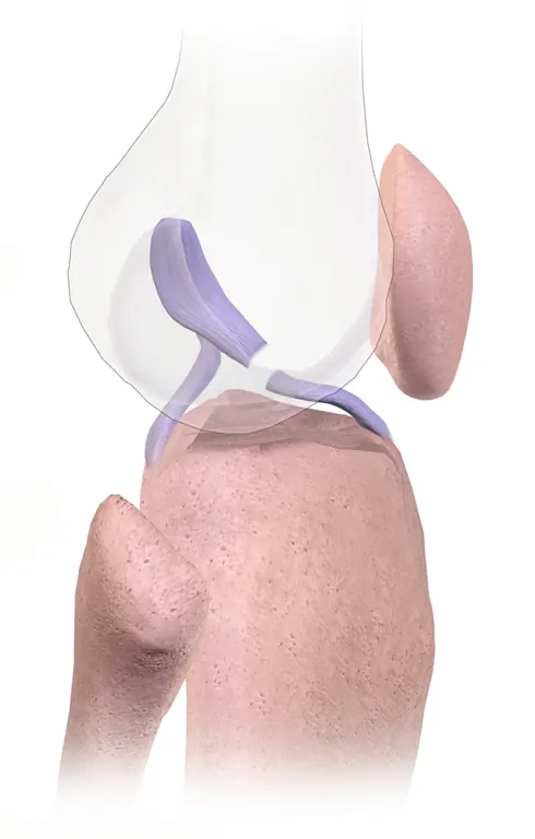
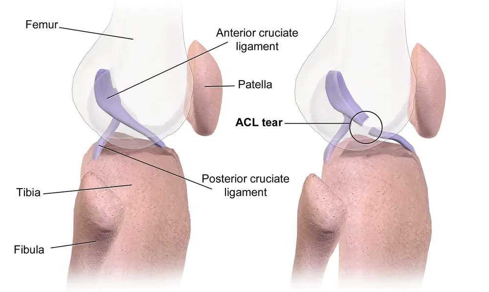
An anterior cruciate ligament (ACL) tear is an injury to one of the four main ligaments that stabilise the knee. The ACL runs diagonally through the middle of the knee and stops the shin bone sliding forwards and rotating too much under the thigh bone.
ACL tears usually happen during sports that involve sudden stops, changes of direction, pivoting or awkward landings, such as football, basketball, netball, rugby and skiing. People often hear or feel a pop, followed by rapid swelling and a feeling that the knee is unstable or gives way. Some people manage well with physiotherapy alone, while others, especially those returning to pivoting sports, need reconstruction surgery followed by many months of rehabilitation.
When to get urgent help
Seek urgent care for a knee that is very swollen and painful after injury, cannot bear weight, is locked and cannot straighten, looks deformed, or has numbness or coldness below the knee.
Causes and risk factors
- Sudden change of direction or pivoting with the foot planted
- Landing awkwardly from a jump, especially with the knee turned inwards
- Sudden deceleration
- Direct blow to the knee, as in tackles or collisions
- Skiing falls
Risk factors include female sex (two to eight times higher risk in some sports, due to anatomy, hormones and movement patterns), previous ACL injury, poor neuromuscular control, playing surfaces and footwear with high grip, and fatigue.
How it develops (pathophysiology)
Normal knee stability
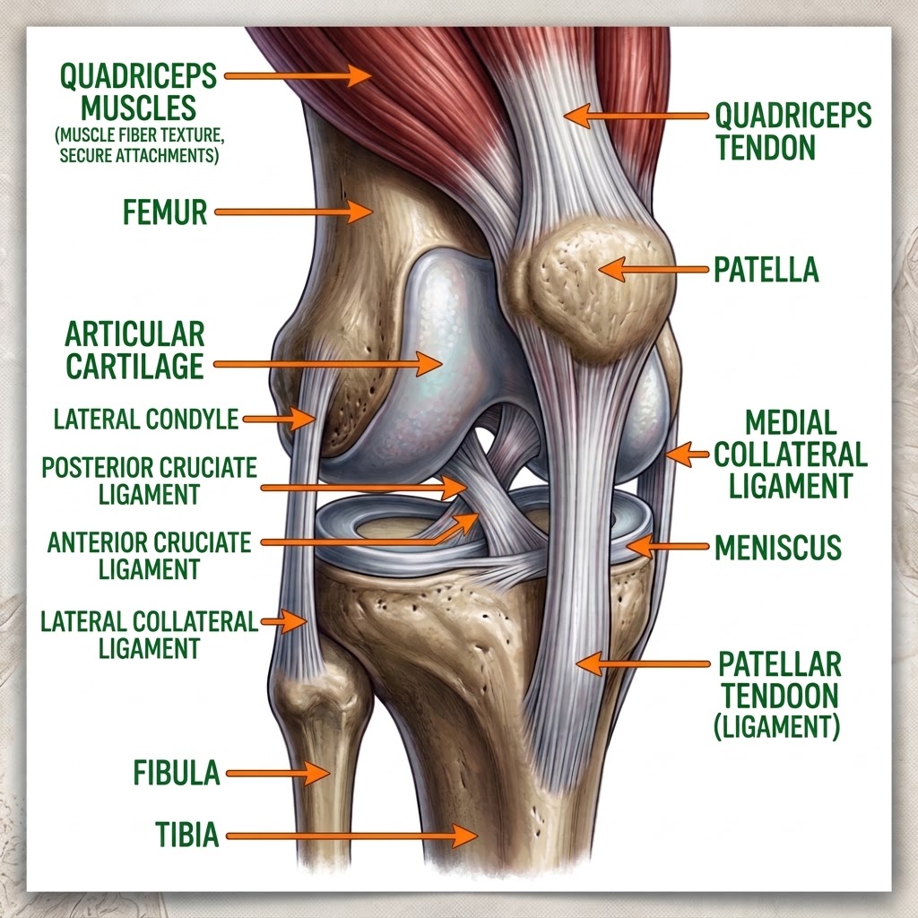
The knee is a hinge joint between the femur (thigh bone) and tibia (shin bone), with the patella (kneecap) in front. Its bony surfaces are fairly flat, so stability depends on ligaments, muscles and the menisci (cartilage cushions):
- The ACL attaches from the front of the tibia to the back of the femur inside the joint. It prevents the tibia moving forwards and controls rotation.
- The posterior cruciate ligament (PCL) prevents the tibia moving backwards.
- The medial and lateral collateral ligaments resist sideways forces.
The ACL is made of collagen bundles and contains nerve endings that sense joint position (proprioception), helping muscles respond quickly.
What goes wrong
- Injury mechanism: most ACL tears are non-contact. When the foot is planted and the knee is slightly bent and turned inwards (valgus collapse), with the tibia rotating, the ACL is stretched beyond its strength and ruptures, often in the middle.
- Associated injuries: the same forces can injure the menisci, medial collateral ligament, cartilage and bone (bone bruises), seen in a common combination called the "unhappy triad".
- Bleeding: the ACL has a blood supply, so tearing it causes bleeding into the joint (haemarthrosis), producing rapid swelling within hours.
- Poor natural healing: unlike ligaments outside the joint, the ACL sits within joint fluid, which prevents a stable blood clot forming between the torn ends. As a result, a completely torn ACL rarely heals back together.
- Instability: without the ACL, the tibia can slide and rotate abnormally, especially during pivoting. This causes episodes of giving way, which can further damage the menisci and cartilage.
Why the symptoms happen
- Pop or snap at the time of injury reflects the ligament rupturing.
- Rapid swelling comes from bleeding into the joint.
- Pain and difficulty bearing weight result from the injury and swelling.
- Feeling of instability or giving way, especially when turning, is caused by abnormal movement of the tibia.
- Reduced range of movement reflects swelling and pain.
Long-term consequences
Repeated instability episodes damage the menisci and cartilage. Even with reconstruction, the risk of osteoarthritis is increased, partly due to the initial cartilage damage.
How treatment acts on the mechanism
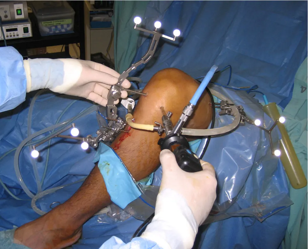
Physiotherapy strengthens the quadriceps and hamstrings and retrains balance and movement, so muscles help control tibial movement. Some people (copers) regain good function this way. ACL reconstruction replaces the torn ligament with a graft (from the patellar tendon, hamstring tendons or quadriceps tendon, or a donor), fixed in tunnels in the bones. The graft gradually remodels into ligament-like tissue over months (ligamentisation), which is why rehabilitation takes 9 to 12 months before return to pivoting sport.
Types
- Grade 1: stretched but intact
- Grade 2: partial tear
- Grade 3: complete tear, the most common
- Isolated ACL tear or combined injuries with menisci or other ligaments
Signs and symptoms
- A pop or snap at the moment of injury
- Rapid swelling within hours
- Pain in the knee
- Difficulty walking or putting weight on the leg
- Feeling that the knee gives way, especially when turning
- Loss of full range of movement
- Tenderness along the joint line (if menisci are also torn)
How it is diagnosed
- History of the injury mechanism
- Examination tests: Lachman test, anterior drawer test and pivot shift test
- X-ray to check for fractures
- MRI scan to confirm the ACL tear and look for meniscus, cartilage and other ligament injuries
Possible complications
- Recurrent instability
- Meniscus tears
- Cartilage damage
- Osteoarthritis in later life
- After surgery: stiffness, graft failure or re-tear, infection, pain at the graft site, blood clots
Goals of treatment
Treatment aims to restore a stable, functional knee, return the person to their chosen activities, and protect the menisci and cartilage.
Medicines
- Pain relief: paracetamol and anti-inflammatories
- Blood clot prevention after surgery if at higher risk
Other treatments
- Initial care: rest, ice, compression, elevation and crutches
- Structured physiotherapy for strength, balance and movement
- ACL reconstruction surgery for people with instability or wanting to return to pivoting sports
- Knee brace in some situations
- Return-to-sport testing before resuming high-risk activity
Living with it and prevention
- Injury prevention programmes (such as FIFA 11+), which train landing, cutting and balance and reduce ACL injuries
- Strengthen hamstrings, quadriceps and hip muscles
- Warm up properly
- Learn safe landing technique: soft landings with knees over toes
- Avoid playing when very fatigued
Monitoring and follow-up
Rehabilitation progress is assessed with strength, hop and functional tests. After reconstruction, follow-up continues for about a year.
Outlook
Most people regain good function. After reconstruction and rehabilitation, many athletes return to sport, though not always at the same level. Long-term arthritis risk is increased.
Special considerations
- Female athletes: higher risk; prevention programmes are especially valuable
- Children and adolescents: growth plates need special surgical techniques
- Older or less active people: often manage well without surgery