
Overview
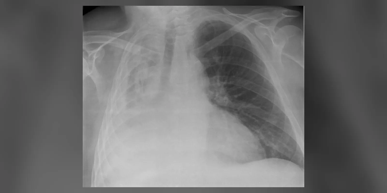
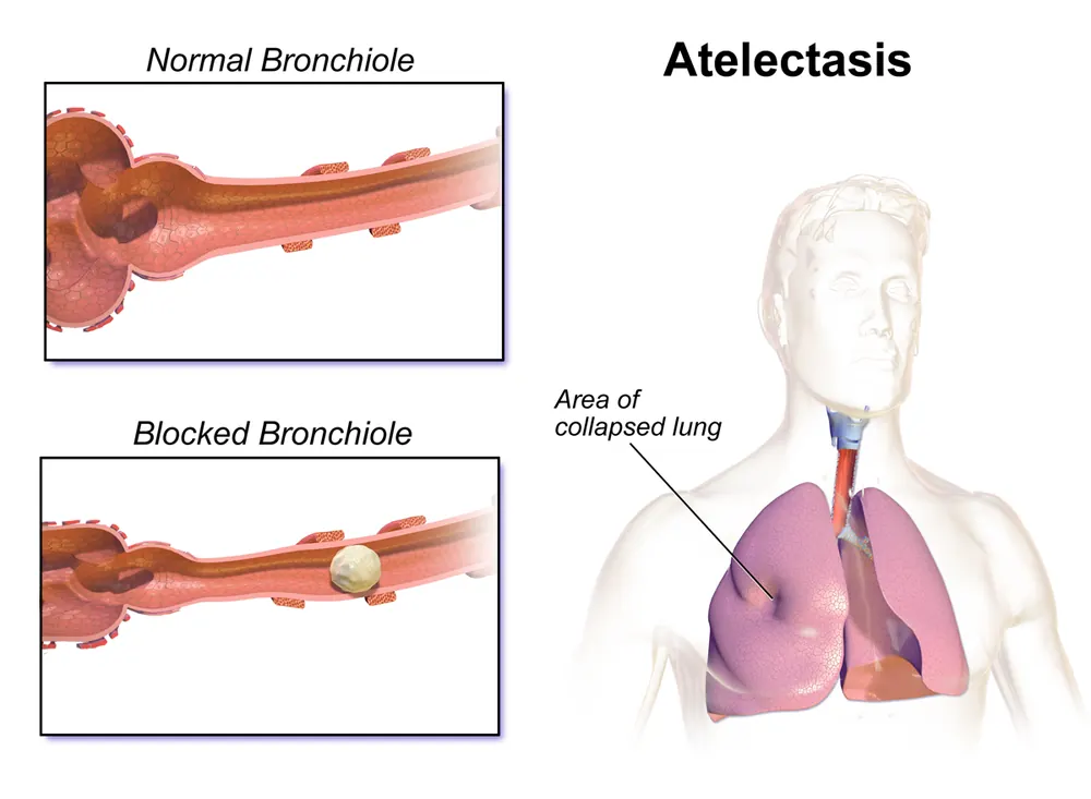
Atelectasis is the collapse or incomplete expansion of part or all of a lung, when the tiny air sacs (alveoli) deflate or fill with fluid. It is not a disease in itself but a sign of another problem.
It most commonly happens after surgery, especially operations on the chest or abdomen under general anaesthetic, when shallow breathing and lying still allow parts of the lungs to collapse. It can also be caused by blockage of an airway by mucus, a tumour or an inhaled object, or by pressure on the lung from fluid, air or a mass. Small areas of atelectasis often cause no symptoms and resolve with deep breathing and movement, while larger collapses can cause breathlessness and low oxygen levels.
When to get urgent help
Seek urgent help for sudden or severe breathlessness, chest pain, blue lips, high fever after surgery, or a child choking or coughing suddenly after possibly inhaling an object.
Causes and risk factors
Obstructive (resorptive) atelectasis, from blocked airways:
- Mucus plugs, common after surgery, in asthma or cystic fibrosis
- Inhaled foreign objects, especially in children
- Tumours inside the airways
- Blood clots
Non-obstructive atelectasis:
- After surgery and anaesthesia: shallow breathing, pain, lying flat
- Compression: pleural effusion, pneumothorax, enlarged heart, tumours
- Lack of surfactant: in premature babies (respiratory distress syndrome)
- Scarring of the lung (cicatricial atelectasis)
- Weak breathing muscles: neuromuscular diseases, spinal cord injury
Risk factors include obesity, smoking, lung disease, older age, and abdominal or chest surgery.
How it develops (pathophysiology)
Normal lung inflation
The lungs stay expanded because the chest wall pulls outward and the lungs naturally recoil inward, creating negative pressure in the space between the lung and chest wall (pleural space). Surfactant, a substance made by cells lining the alveoli, reduces surface tension and stops the tiny air sacs from collapsing when we breathe out. Regular deep breaths and sighs reopen any small areas that begin to collapse.
What goes wrong
Obstructive atelectasis:
- An airway becomes blocked, for example by mucus.
- Air already in the alveoli beyond the blockage is gradually absorbed into the blood.
- With no fresh air entering, the alveoli deflate and collapse.
Post-operative atelectasis:
- General anaesthesia relaxes the diaphragm and chest muscles, reducing lung volume.
- High oxygen concentrations during anaesthesia speed up absorption of gas from poorly ventilated alveoli.
- After surgery, pain limits deep breathing and coughing, so mucus builds up and small airways close.
- Lying flat further reduces lung volume, especially at the bases.
Compressive atelectasis:
- Fluid, air, blood or a mass in the chest presses on the lung, squeezing alveoli closed.
Surfactant deficiency:
- Without surfactant, surface tension causes alveoli to collapse, as in premature babies or acute lung injury.
Consequences
- Ventilation-perfusion mismatch: blood still flows through collapsed lung but does not pick up oxygen, causing low oxygen levels (a shunt).
- Infection: trapped mucus provides a breeding ground for bacteria, leading to pneumonia.
- Lung function: large collapses reduce the lung volume available for breathing.
Why the symptoms happen
- Breathlessness and fast breathing result from low oxygen and reduced lung volume.
- Cough reflects the underlying cause, such as mucus.
- Chest pain may occur with an underlying cause such as pneumothorax.
- Fever after surgery may reflect atelectasis or developing infection.
- Reduced breath sounds occur over the collapsed area.
How treatment acts on the mechanism
Deep breathing exercises, incentive spirometry and coughing reopen collapsed alveoli and clear mucus. Early mobilisation after surgery increases lung volume. Chest physiotherapy helps clear secretions. Bronchoscopy removes mucus plugs or foreign bodies. Draining fluid or air relieves compression. Positive pressure (CPAP) keeps alveoli open. Treating the underlying cause, such as a tumour, restores airway patency.
Types
- Obstructive (resorptive) atelectasis
- Compressive atelectasis
- Post-operative atelectasis
- Adhesive atelectasis (surfactant deficiency)
- Cicatricial (scarring) atelectasis
- Rounded atelectasis, often linked to asbestos exposure
- By extent: subsegmental, segmental, lobar or whole lung
Signs and symptoms
- Often no symptoms if small
- Breathlessness
- Rapid, shallow breathing
- Cough
- Low-grade fever, especially after surgery
- Chest discomfort
- Blue lips or skin if oxygen levels are very low
How it is diagnosed
- Examination: reduced breath sounds and dullness over the affected area
- Oxygen saturation
- Chest X-ray: shows collapse and shift of structures towards the affected side
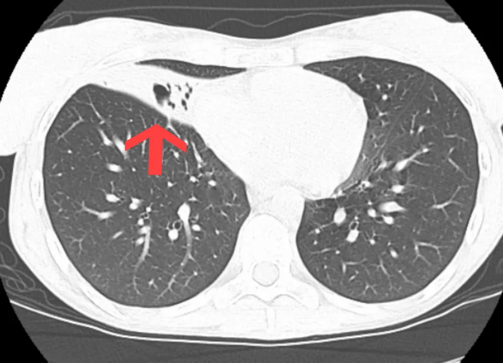
- CT scan to identify the cause, such as a tumour
- Bronchoscopy to look for and remove blockages
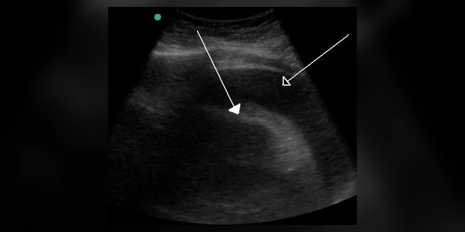
- Ultrasound to identify pleural fluid
Possible complications
- Low oxygen levels
- Pneumonia
- Respiratory failure in severe cases
- Bronchiectasis from long-term collapse
Goals of treatment
Treatment aims to re-expand the lung, restore oxygen levels, clear secretions and treat the underlying cause.
Medicines
- Bronchodilators if airway narrowing contributes
- Mucus-thinning nebulisers such as saline
- Antibiotics if infection develops
- Pain relief after surgery so that deep breathing and coughing are possible
Other treatments
- Deep breathing and coughing exercises
- Incentive spirometer
- Early walking after surgery
- Chest physiotherapy and postural drainage
- Positioning: sitting up, lying on the unaffected side
- CPAP or other positive pressure support
- Bronchoscopy to remove plugs or foreign bodies
- Chest drain for fluid or air
- Treatment of tumours
Living with it and prevention
- Stop smoking before surgery
- Practise deep breathing exercises before and after surgery
- Move around as soon as possible after surgery
- Use pain relief so you can breathe deeply and cough
- Keep children away from small objects they could inhale
Monitoring and follow-up
Oxygen levels and chest X-rays may be repeated to check re-expansion. Persistent or unexplained collapse needs investigation for an underlying cause such as cancer.
Outlook
Most post-operative atelectasis resolves within days with breathing exercises and mobilisation. The outlook in other cases depends on the underlying cause.
Special considerations
- Premature babies: surfactant deficiency causes respiratory distress syndrome; treated with surfactant and breathing support
- Children: inhaled foreign bodies are a common cause
- People with obesity or neuromuscular disease: higher risk