
Overview
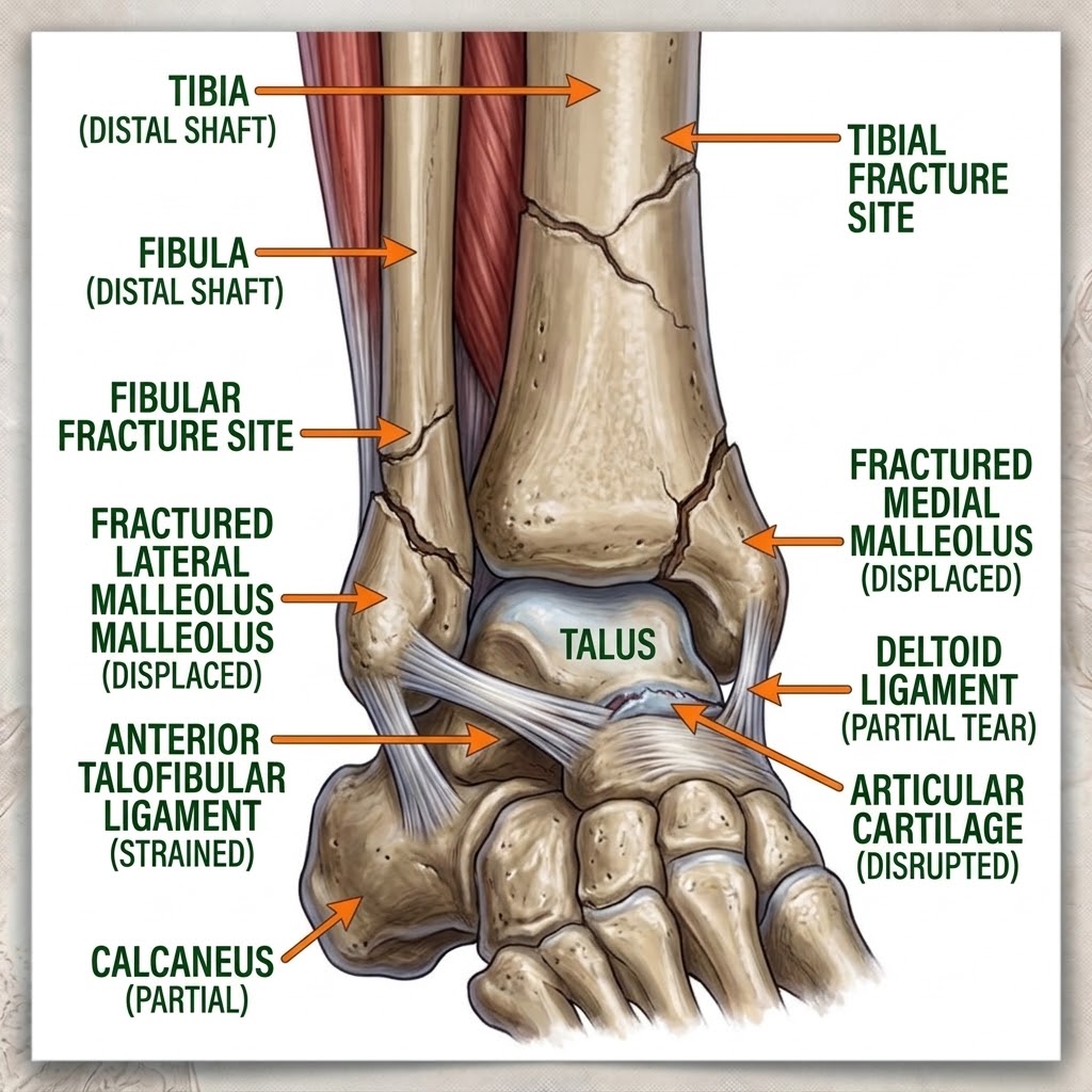
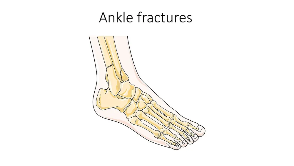
An ankle fracture is a break in one or more of the bones that make up the ankle joint: the lower ends of the tibia (shin bone) and fibula (the thinner outer leg bone), and sometimes the talus, the bone beneath them. Ankle fractures are among the most common bone injuries and can happen at any age, from sports injuries in young people to falls in older adults with weaker bones.
Fractures range from small cracks in one bone that heal with a supportive boot to complex breaks of several bones with damage to the ligaments, making the joint unstable. Stable fractures are usually treated without surgery, while unstable fractures often need an operation to realign and fix the bones. Most people recover well, although healing and rehabilitation can take several months.
When to get urgent help
Seek emergency care for a deformed ankle, bone through the skin, a cold, pale or numb foot, severe pain not controlled by painkillers, or calf pain and swelling or breathlessness after injury (possible blood clot).
Causes and risk factors
- Twisting injuries, such as rolling the ankle inwards or outwards on uneven ground
- Falls, including from height
- Sports injuries, especially football, basketball, skiing and running
- Road traffic accidents
- Direct blows to the ankle
Risk factors include osteoporosis, older age, obesity, diabetes with neuropathy, smoking, previous ankle injuries and wearing unsupportive footwear.
How it develops (pathophysiology)
Normal structure of the ankle
The ankle joint is a hinge formed by the talus sitting in a socket called the mortise, made by the lower tibia and its inner bump (the medial malleolus), the back edge of the tibia (the posterior malleolus), and the lower fibula (the lateral malleolus). The bones are held together by strong ligaments:
- The deltoid ligament on the inner side
- The lateral ligaments on the outer side
- The syndesmosis, a set of ligaments binding the tibia and fibula together just above the joint
This arrangement keeps the talus tightly centred, allowing smooth up-and-down movement while resisting twisting.
What goes wrong
Most ankle fractures happen when the foot is planted and the leg rotates, or the foot twists under the body. The pattern depends on the position of the foot and the direction of force:
- Supination-external rotation, the most common mechanism, causes a spiral fracture of the fibula at the level of the joint, sometimes with ligament or bone injury on the inner side.
- Pronation injuries can cause a fibula fracture higher up the leg and a break or tear on the inner side.
- When both sides of the ankle are broken (bimalleolar) or all three malleoli (trimalleolar), the ring of bone and ligaments around the talus is disrupted.
The key concept is stability. If the bones or ligaments on only one side are injured, the talus stays centred and the joint is stable. If both sides are injured or the syndesmosis is torn, the talus can shift. Even a 1 millimetre shift of the talus reduces the contact area of the joint by about 40 percent, concentrating force on cartilage and greatly increasing the risk of arthritis.
Healing
Bone heals in stages: a blood clot forms around the break, inflammatory cells clear debris, a soft callus of cartilage and fibrous tissue bridges the gap within weeks, then hard bone replaces it over two to three months, followed by remodelling over a year or more. Smoking, diabetes and poor circulation slow this process.
Why the symptoms happen
- Severe pain results from bone and soft tissue injury and pressure from bleeding.
- Swelling and bruising are caused by bleeding from bone and torn vessels.
- Inability to bear weight occurs because the joint is unstable and painful.
- Deformity indicates displacement or dislocation.
- Numbness or a cold foot suggests nerve or blood vessel compression, an emergency.
How treatment acts on the mechanism
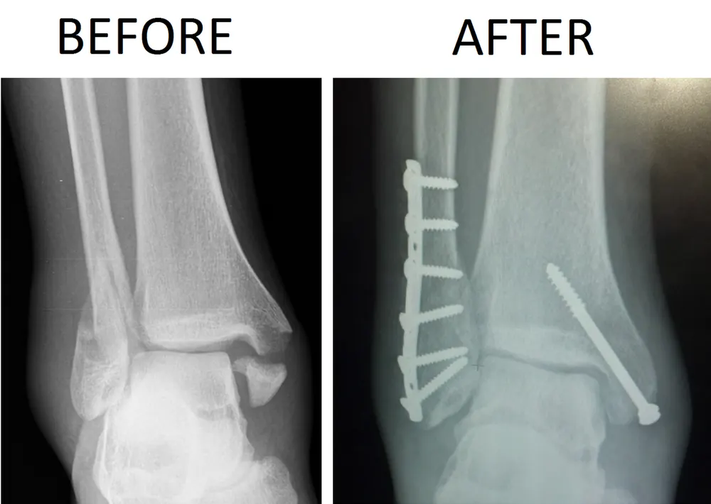
Immobilisation in a boot or cast holds stable fractures in position while callus forms. Reduction (realigning the bones) restores the mortise. Open reduction and internal fixation (ORIF) uses plates and screws to hold unstable fractures precisely, recreating a congruent joint to lower arthritis risk. Syndesmosis screws or suture devices stabilise torn ligaments between tibia and fibula. Rehabilitation restores movement, strength and balance.
Types
- Lateral malleolus fracture: break of the fibula only; often stable
- Medial malleolus fracture
- Bimalleolar fracture: both sides
- Trimalleolar fracture: both sides plus the back of the tibia
- Pilon fracture: high-energy break of the tibial weight-bearing surface
- Syndesmotic injuries
- Classified by Weber (A, B, C, based on fibula fracture level) or Lauge-Hansen systems
Signs and symptoms
- Immediate severe pain
- Swelling and bruising
- Tenderness over the bone
- Difficulty or inability to put weight on the foot
- Visible deformity in some cases
- A crack or snap at the time of injury
- Numbness, tingling or a pale foot if nerves or vessels are affected
How it is diagnosed
- Examination, using the Ottawa ankle rules to decide whether an X-ray is needed (tenderness over specific bone areas or inability to walk four steps)
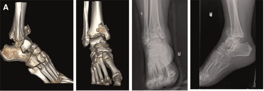
- X-rays of the ankle, and sometimes the full length of the lower leg
- Stress X-rays or weight-bearing X-rays to assess stability
- CT scan for complex fractures
- MRI for ligament or cartilage injury
Possible complications
- Poor healing (delayed union or non-union)
- Healing in the wrong position (malunion)
- Post-traumatic arthritis
- Stiffness and long-term pain
- Blood clots in the legs (DVT) or lungs
- Wound infection after surgery
- Compartment syndrome
- Complex regional pain syndrome
- Skin breakdown in people with diabetes
Goals of treatment
Treatment aims to restore a stable, well-aligned ankle joint, allow bone healing, relieve pain, and return the person to normal walking and activities.
Medicines
- Pain relief: paracetamol, anti-inflammatories or short courses of stronger painkillers
- Blood clot prevention (anticoagulants) for people at higher risk while immobilised
- Antibiotics for open fractures
Other treatments
- Rest, ice and elevation to reduce swelling
- Walking boot or cast for stable fractures, often allowing weight-bearing as tolerated
- Closed reduction under sedation for displaced fractures
- Surgery (ORIF) for unstable or displaced fractures
- External fixator for severe swelling or complex injuries before definitive surgery
- Physiotherapy for movement, strength and balance
Living with it and prevention
- Wear supportive footwear and take care on uneven ground
- Strengthen leg muscles and improve balance, especially in older adults
- Treat osteoporosis
- Use ankle supports for high-risk sports if advised
During recovery:
- Keep the leg raised to reduce swelling
- Do not smoke, as it slows bone healing
- Follow weight-bearing advice
- Do prescribed exercises
Monitoring and follow-up
Follow-up X-rays check alignment and healing, typically at one, two and six weeks. Physiotherapy continues until walking and strength are restored. Most people return to normal activities in three to four months.
Outlook
Most people recover well. Stable fractures usually heal in about six weeks. Unstable fractures treated surgically also heal well, but some people develop stiffness or arthritis.
Special considerations
- Older adults: fractures may be linked to osteoporosis and need bone health assessment
- People with diabetes: higher risk of wound and healing problems; careful follow-up is essential
- Children: injuries may involve growth plates and need specialist care