
Overview
An ankle sprain is an injury to the ligaments, the strong bands of tissue that connect the bones of the ankle. It usually happens when the foot rolls or twists awkwardly, stretching or tearing one or more ligaments. Ankle sprains are one of the most common injuries, particularly in sports involving jumping, running and changing direction, but they can also happen while walking on uneven ground.
Most sprains affect the ligaments on the outer side of the ankle. Symptoms include pain, swelling, bruising and difficulty walking. Most ankle sprains heal well within a few weeks with early movement, gradual return to activity and exercises to restore strength and balance. Without proper rehabilitation, some people develop repeated sprains and long-term ankle instability.
When to get urgent help
Seek urgent care for inability to walk four steps, severe pain, deformity, numbness, a cold foot, or tenderness over the bones of the ankle or foot, which may indicate a fracture.
Causes and risk factors
- Rolling the ankle inwards (inversion), the most common mechanism
- Sports: basketball, volleyball, football, netball and trail running
- Uneven surfaces, steps and kerbs
- Landing awkwardly after a jump
- Unsupportive footwear or high heels
Risk factors include previous ankle sprains, poor balance, weak ankle muscles, reduced ankle flexibility and fatigue.
How it develops (pathophysiology)
Normal ankle stability
The ankle is held stable by bone shape and ligaments:
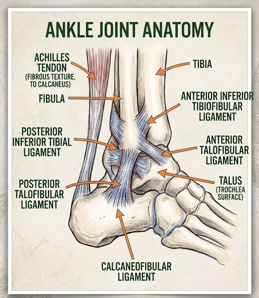
- Lateral ligaments on the outside: the anterior talofibular ligament (ATFL), calcaneofibular ligament (CFL) and posterior talofibular ligament (PTFL)
- The deltoid ligament on the inside, which is very strong
- The syndesmosis, ligaments binding the tibia and fibula together
Ligaments are made of tightly packed collagen fibres. They resist excessive movement and contain nerve endings that sense joint position (proprioception), helping muscles react quickly to prevent injury.
What goes wrong
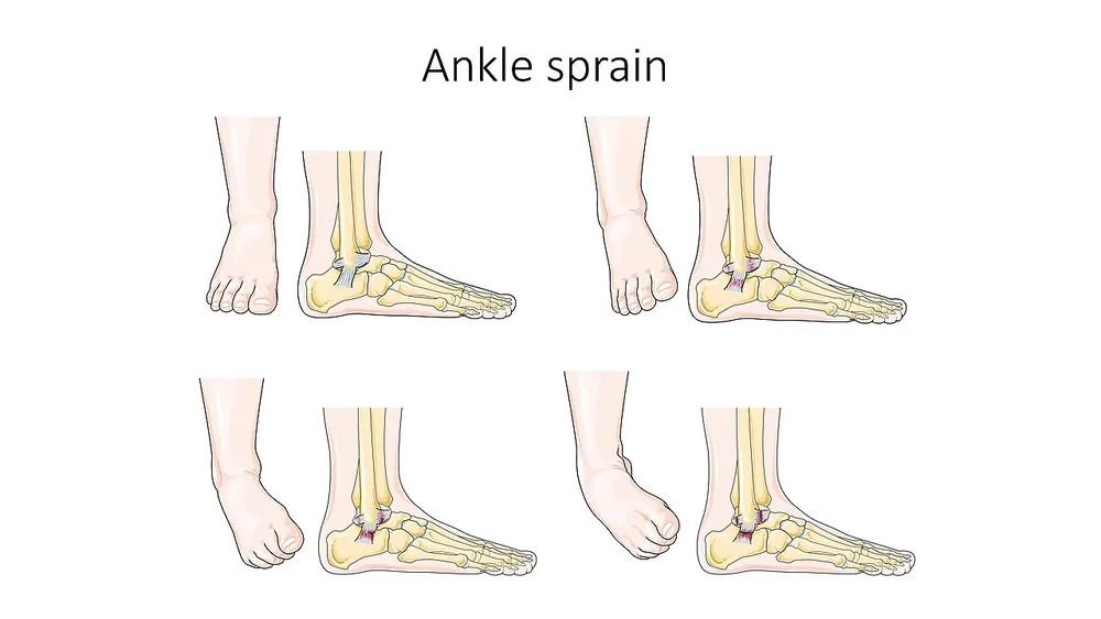
- Injury mechanism: when the foot rolls inwards while pointed down, the ATFL is under the most strain and is the first to tear. With greater force, the CFL tears too. The PTFL is rarely injured.
- Tissue damage: collagen fibres are stretched (grade 1), partly torn (grade 2) or completely torn (grade 3).
- Inflammation: bleeding and release of inflammatory mediators cause swelling, warmth and pain over the first few days.
- Repair: fibroblasts lay down new collagen, initially disorganised type III collagen, which gradually remodels into stronger type I collagen over weeks to months. The repaired ligament may remain slightly longer and weaker than before.
- Proprioception loss: damaged nerve endings reduce the ankle's ability to sense position, so muscles react more slowly to future twists.
A high ankle sprain involves the syndesmosis, typically from the foot being forced outwards and rotated, and takes longer to heal.
Why the symptoms happen
- Pain and tenderness over the outer ankle come from torn ligament fibres and inflammation.
- Swelling and bruising result from bleeding and fluid leakage.
- Difficulty walking reflects pain and instability.
- Feeling of the ankle giving way occurs with severe tears or poor proprioception.
Chronic ankle instability
About one in five people who sprain their ankle develop chronic ankle instability, with repeated sprains and a feeling of looseness. This is caused by stretched ligaments (mechanical instability) and impaired proprioception and muscle control (functional instability).
How treatment acts on the mechanism
Early protected movement encourages collagen fibres to align properly as they heal. Compression and elevation limit swelling. Bracing protects the healing ligament. Balance and strengthening exercises retrain proprioception and the muscles (especially the peroneal muscles on the outside of the leg) that protect the ankle, reducing recurrence. Surgery repairs or reconstructs ligaments in chronic instability.
Types
- Grade 1 (mild): stretching with minimal tearing
- Grade 2 (moderate): partial tear, some instability
- Grade 3 (severe): complete tear, marked instability
- Lateral ankle sprain: most common
- Medial ankle sprain: less common
- High ankle (syndesmotic) sprain
Signs and symptoms
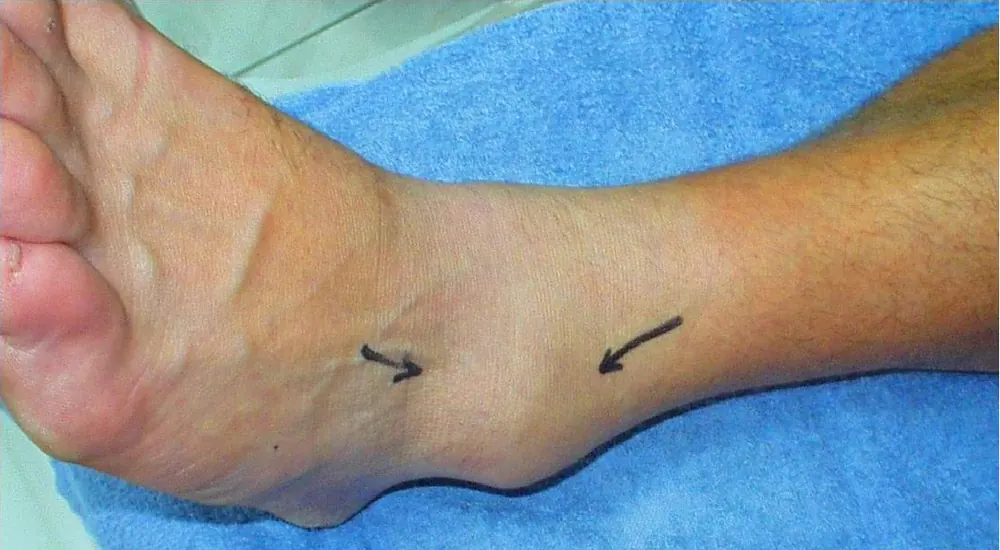
- Pain on the outside of the ankle
- Swelling
- Bruising, which may spread to the foot
- Tenderness when pressing on the ligament
- Difficulty putting weight on the foot
- Stiffness
- A feeling that the ankle is unstable
How it is diagnosed
- Examination of the ankle and foot
- Ottawa ankle rules to decide if an X-ray is needed to exclude a fracture
- Anterior drawer and talar tilt tests for ligament laxity, often done a few days after injury
- Ultrasound or MRI for persistent symptoms or suspected high ankle sprain
Possible complications
- Chronic ankle instability and repeated sprains
- Persistent pain or swelling
- Cartilage injury to the talus
- Peroneal tendon injury
- Arthritis in the long term
Goals of treatment
Treatment aims to reduce pain and swelling, restore movement, strength and balance, and prevent recurrence.
Medicines
- Paracetamol for pain
- Anti-inflammatory medicines (such as ibuprofen tablets or gel) after the first 48 hours, if suitable
Other treatments
- PEACE and LOVE approach: Protect, Elevate, Avoid anti-inflammatories early, Compress, Educate; then Load, Optimism, Vascularisation (pain-free cardiovascular exercise), Exercise
- Ice for short periods may relieve pain
- Ankle brace or support
- Crutches briefly if needed
- Physiotherapy: range-of-motion, strengthening and balance exercises (such as single-leg standing and wobble board)
- Gradual return to sport
- Surgery for chronic instability that does not improve with rehabilitation
Living with it and prevention
- Warm up before exercise
- Wear supportive footwear
- Do balance and ankle strengthening exercises regularly
- Use a brace or taping for sports after a previous sprain
- Take care on uneven ground
Monitoring and follow-up
Most mild sprains improve within two weeks, and moderate sprains within six weeks. Persistent pain, swelling or instability beyond six weeks should be reviewed.
Outlook
Most people recover fully. Completing rehabilitation reduces the risk of repeat sprains. A minority develop chronic instability.
Special considerations
- Athletes: sport-specific rehabilitation and bracing reduce recurrence
- Children: ligaments are often stronger than growth plates, so a "sprain" may be a growth plate fracture
- Older adults: consider fracture and falls risk