
Overview
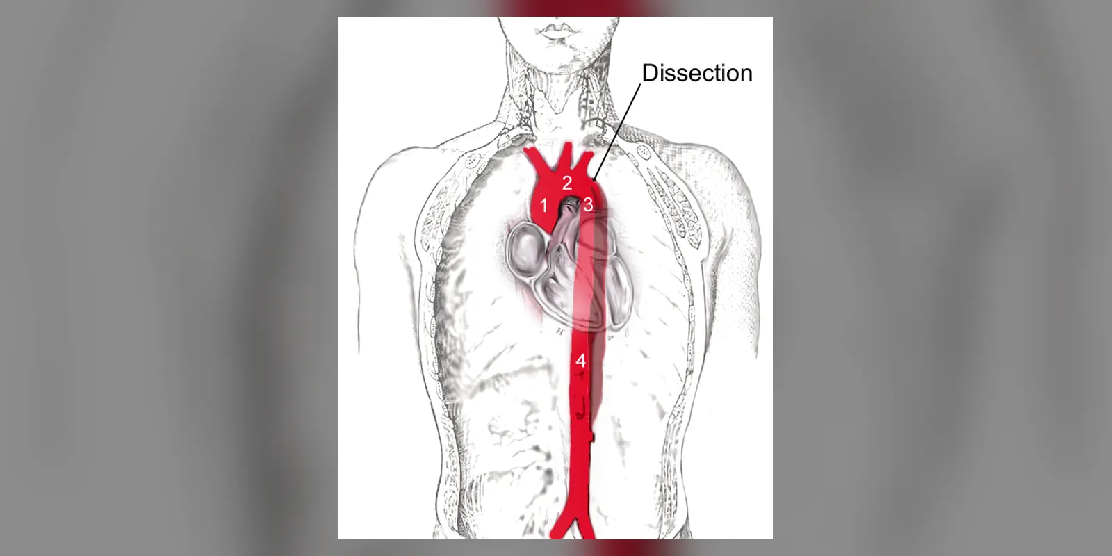
Aortic dissection is a medical emergency in which a tear develops in the inner layer of the aorta, the body's main artery. Blood surges through the tear and forces apart the layers of the artery wall, creating a false channel. This can block blood flow to organs, cause the aorta to burst, or damage the aortic valve and heart.
The classic symptom is sudden, severe, tearing or ripping pain in the chest or back. Aortic dissection is uncommon but often fatal without rapid treatment. It is most common in men aged 60 to 80 with high blood pressure, but it can occur in younger people with inherited connective tissue disorders such as Marfan syndrome. Treatment involves urgent blood pressure control and often emergency surgery.
When to get urgent help
Call emergency services immediately for sudden severe chest or back pain, especially if tearing in nature or accompanied by fainting, breathlessness, weakness or stroke symptoms.
Causes and risk factors
- High blood pressure: the most important risk factor
- Atherosclerosis
- Older age
- Inherited connective tissue disorders: Marfan syndrome, Ehlers-Danlos syndrome (vascular type), Loeys-Dietz syndrome, Turner syndrome
- Bicuspid aortic valve and coarctation of the aorta
- Pre-existing aortic aneurysm
- Cocaine or amphetamine use
- Pregnancy, especially with underlying conditions
- Heavy weightlifting
- Injury to the chest
- Previous heart surgery or procedures on the aorta
How it develops (pathophysiology)
Normal structure of the aorta
The aortic wall has three layers:
- The intima, a thin inner lining
- The media, a thick middle layer of elastic fibres and smooth muscle that gives strength and elasticity
- The adventitia, an outer layer of connective tissue
With each heartbeat, the aorta stretches and recoils, absorbing the force of blood ejected by the heart.
What goes wrong
- Weakening of the media: years of high blood pressure, ageing or inherited conditions damage elastic fibres and smooth muscle cells, a process called medial degeneration. In Marfan syndrome, faulty fibrillin-1 and excessive TGF-beta signalling weaken the wall from birth.
- Intimal tear: a tear forms in the intima, usually where stress is highest, just above the aortic valve or just beyond the arteries to the left arm.
- Propagation: blood under high pressure enters the media and splits it, creating a false lumen that can extend along the aorta in either direction.
- Consequences: - Branch artery blockage: the false lumen can compress or tear the origins of arteries to the heart, brain, spinal cord, gut, kidneys and legs, causing stroke, heart attack, paralysis or organ damage - Rupture: the thin outer wall can burst into the chest, abdomen or the sac around the heart (cardiac tamponade) - Aortic valve regurgitation: dissection near the heart distorts the valve - Aneurysm: in survivors, the weakened wall may enlarge over time
Intramural haematoma (bleeding within the wall without a clear tear) and penetrating aortic ulcer are related conditions.
Why the symptoms happen
- Sudden, severe, tearing pain results from the splitting of the aortic wall, which is rich in nerve endings. The pain may move as the dissection extends.
- Chest pain suggests the ascending aorta; back pain between the shoulder blades suggests the descending aorta.
- Unequal pulses or blood pressure between the arms occur when arm arteries are affected.
- Fainting, stroke symptoms or limb weakness result from blocked branches.
- Low blood pressure and shock indicate rupture or tamponade.
How treatment acts on the mechanism
Beta-blockers lower heart rate and the force of each heartbeat, reducing the shearing force on the aortic wall and slowing propagation. Blood pressure lowering reduces stress. Emergency surgery for ascending (type A) dissection replaces the damaged aorta with a graft and repairs or replaces the aortic valve. Endovascular stent grafts seal the entry tear in complicated descending (type B) dissections, redirecting blood into the true lumen.
Types
- Stanford type A: involves the ascending aorta; usually needs emergency surgery
- Stanford type B: involves only the descending aorta; often managed with medicines unless complicated
- DeBakey types I, II and III
- Acute (less than two weeks), subacute and chronic
Signs and symptoms
- Sudden, severe chest or upper back pain, often described as tearing, ripping or stabbing
- Pain that moves to the neck, jaw, abdomen or legs
- Sweating and paleness
- Breathlessness
- Fainting
- Weakness or numbness on one side, difficulty speaking or vision loss (stroke symptoms)
- Weak or absent pulse in one arm or leg
- Abdominal pain or leg pain if branches to the gut or legs are blocked
How it is diagnosed
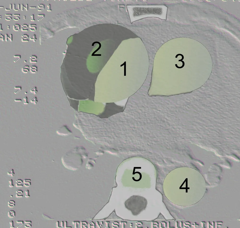
- CT angiography, the main test
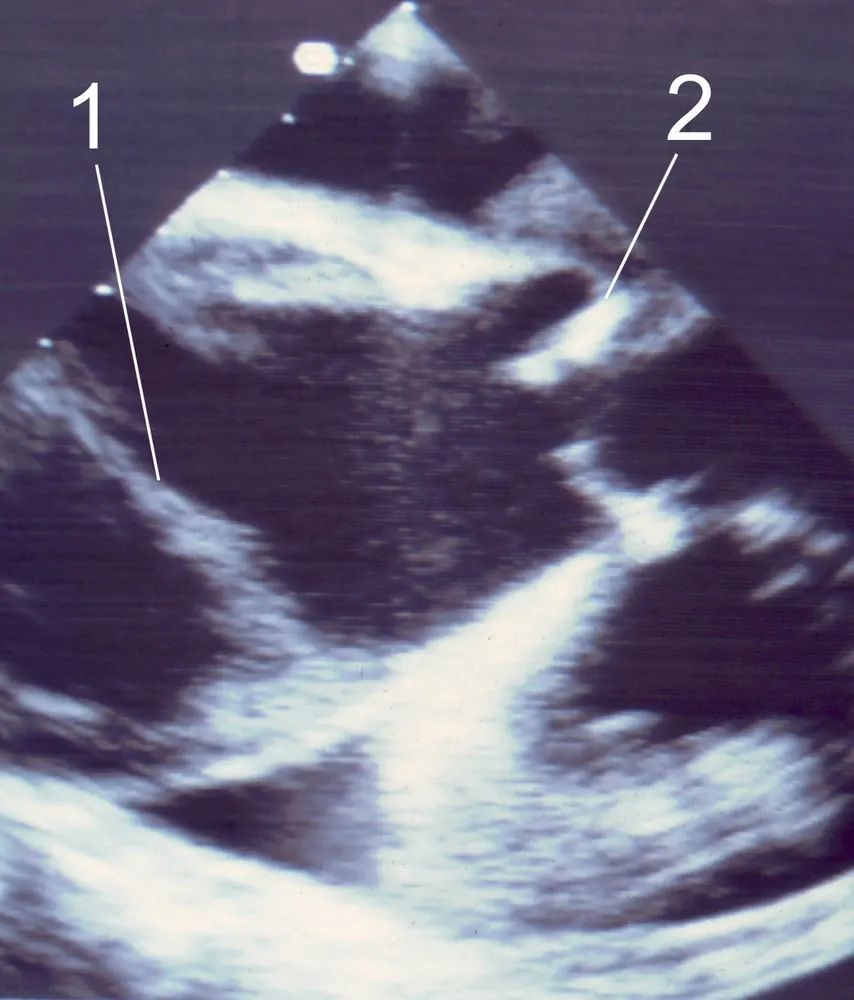
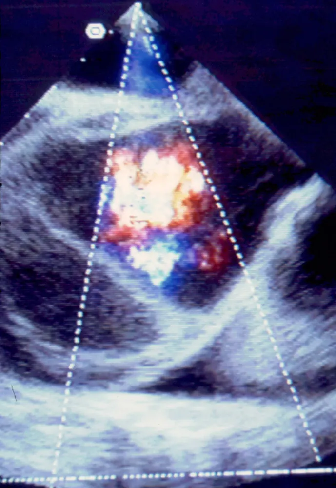
- Transoesophageal echocardiogram in unstable patients
- MRI angiography for stable patients or follow-up
- ECG and blood tests, including D-dimer and troponin, to help distinguish from heart attack
- Blood pressure in both arms
Possible complications
- Aortic rupture and death
- Cardiac tamponade
- Heart attack
- Stroke
- Paraplegia from spinal cord ischaemia
- Kidney failure
- Bowel ischaemia
- Aortic valve regurgitation and heart failure
- Late aneurysm formation
Goals of treatment
Treatment aims to stop the dissection spreading, prevent rupture, restore blood flow to organs, and provide long-term protection of the aorta.
Medicines
- Intravenous beta-blockers (such as labetalol or esmolol) to lower heart rate and blood pressure
- Additional blood pressure medicines if needed
- Strong pain relief
- Long-term blood pressure control
Other treatments
- Emergency open surgery for type A dissection
- Thoracic endovascular aortic repair (TEVAR) for complicated type B dissection
- Intensive care monitoring
Living with it and prevention
- Control blood pressure
- Do not use cocaine or amphetamines
- Avoid heavy weightlifting if at risk
- People with Marfan syndrome or related conditions need regular aortic imaging and may have preventive surgery
- Family screening for inherited aortic disease
Monitoring and follow-up
After dissection, people need lifelong blood pressure control and regular CT or MRI scans to check for aneurysm growth.
Outlook
Untreated type A dissection has a very high death rate in the first days. With prompt surgery, most people survive. Type B dissection has better short-term survival with medical treatment, but long-term monitoring is essential.
Special considerations
- Pregnancy: higher risk in women with Marfan syndrome or other aortic diseases
- Young people with dissection: should be assessed for inherited conditions
- Family members: genetic testing and imaging may be offered