
Overview
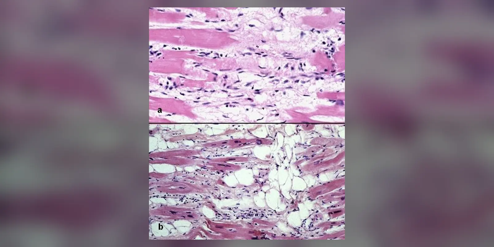
Arrhythmogenic cardiomyopathy (ACM), often called arrhythmogenic right ventricular cardiomyopathy (ARVC), is an inherited heart muscle disease. Over time, healthy heart muscle, most often in the right ventricle, is gradually replaced by fat and scar tissue. This disrupts the heart's electrical signals and can cause dangerous fast heart rhythms from the ventricles.
ACM usually becomes apparent in adolescence or young adulthood and is an important cause of sudden cardiac death in young people and athletes. Symptoms include palpitations, dizziness and fainting, particularly during exercise. Treatment focuses on preventing dangerous rhythms, through avoiding intense exercise, medicines, and in many cases an implantable cardioverter-defibrillator (ICD), and on screening family members.
Because many people with a gene change have no symptoms, the condition is often found through family screening after a relative is diagnosed or dies suddenly. Early recognition allows simple measures, such as avoiding intense exercise, to make a real difference.
When to get urgent help
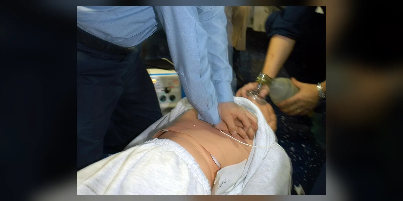
Call emergency services for fainting, chest pain, palpitations with dizziness, or collapse. If someone collapses and is not breathing normally, start CPR and use a defibrillator (AED).
Causes and risk factors
- Inherited gene changes affecting desmosomes, structures that link heart muscle cells together, especially the PKP2 gene; others include DSP, DSG2, DSC2 and JUP
- Usually inherited in an autosomal dominant pattern (50 percent chance of passing it on), with variable severity
- Rarer recessive forms (Naxos disease, Carvajal syndrome) also affect hair and skin
- Endurance exercise accelerates disease in people with gene changes
- Some cases have no identified gene
How it develops (pathophysiology)
Normal heart muscle structure
Heart muscle cells (cardiomyocytes) are joined end to end by intercalated discs. These contain three types of connection:
- Desmosomes: mechanical anchors that hold cells together under the stress of contraction
- Gap junctions: channels that let electrical signals pass quickly from cell to cell
- Adherens junctions: anchor the internal skeleton
Proper desmosome function is needed for both mechanical strength and normal electrical conduction.
What goes wrong
- Faulty desmosomes: gene changes produce weak or missing desmosome proteins. Under mechanical stress, especially during exercise, cells start to detach from one another.
- Cell damage and death: detached cells are injured and die. The thinner-walled right ventricle, which stretches more, is often affected first.
- Fibrofatty replacement: dead muscle is replaced by fat and scar tissue, starting from the outer layer of the heart wall and moving inwards. Signalling changes (including the Wnt and Hippo pathways) push repair cells towards becoming fat cells.
- Electrical disruption: scar and fat interrupt conduction. Gap junctions are reduced, so electrical signals travel slowly and unevenly, creating circuits where signals loop around scar (re-entry), causing ventricular tachycardia.
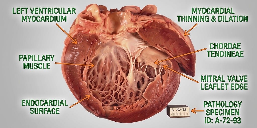
- Structural changes: the right ventricle enlarges, thins and develops bulges (aneurysms). In some people, the left ventricle is affected (left-dominant ACM), and late disease can cause heart failure.
Inflammation (myocarditis-like episodes) may cause "hot phases" with chest pain and raised troponin.
Why the symptoms happen
- Palpitations come from extra beats or ventricular tachycardia.
- Dizziness and fainting occur when fast rhythms reduce cardiac output.
- Sudden cardiac arrest results from ventricular fibrillation, often during exercise when adrenaline makes damaged tissue more irritable.
- Breathlessness and swollen legs develop when the ventricles weaken.
How treatment acts on the mechanism
Exercise restriction reduces mechanical stress on desmosomes and slows disease progression. Beta-blockers reduce adrenaline-driven arrhythmias. Antiarrhythmic medicines such as sotalol, amiodarone or flecainide help control rhythms. An ICD detects dangerous rhythms and delivers a shock or pacing to restore normal rhythm, preventing sudden death. Catheter ablation destroys tissue forming re-entry circuits.
Types
- Right-dominant ACM (classic ARVC)
- Biventricular ACM
- Left-dominant ACM, often with desmoplakin gene changes
Signs and symptoms
- Palpitations
- Light-headedness or dizziness
- Fainting, especially during or after exercise
- Breathlessness
- Chest pain
- Swollen legs or abdomen in later stages
- Sudden cardiac arrest, sometimes as the first sign
Symptoms often first appear during or shortly after physical exertion, when adrenaline levels are high. Some people have no symptoms for many years.
How it is diagnosed
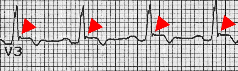
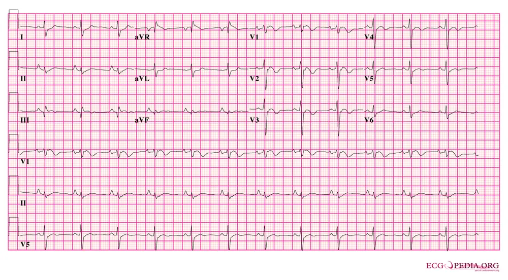
- ECG: may show characteristic changes, such as inverted T waves in leads V1 to V3 and epsilon waves
- Holter or event monitoring for extra beats and arrhythmias
- Echocardiogram
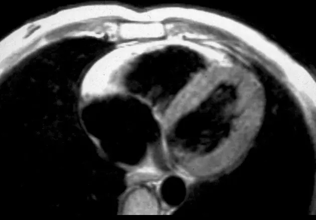
- Cardiac MRI: shows ventricular enlargement, wall motion problems, fat and scar
- Exercise testing
- Genetic testing
- Family history
- Diagnosis uses the Task Force criteria combining these findings
Possible complications
- Ventricular tachycardia
- Sudden cardiac death
- Heart failure
- Blood clots and stroke (less common)
- ICD complications: inappropriate shocks, lead problems
Goals of treatment
Treatment aims to prevent sudden death, control arrhythmias, slow disease progression and support family screening.
Medicines
- Beta-blockers
- Antiarrhythmics: sotalol, amiodarone, flecainide
- Heart failure medicines in advanced disease
- Anticoagulants if clots or atrial fibrillation develop
Other treatments
- Implantable cardioverter-defibrillator (ICD) for people at high risk
- Catheter ablation for recurrent ventricular tachycardia
- Exercise restriction, avoiding competitive and high-intensity endurance sport
- Heart transplantation in severe heart failure or uncontrollable arrhythmia
- Genetic counselling
Living with it and prevention
- Avoid competitive sports and intense endurance exercise if diagnosed or a gene carrier
- Take medicines as prescribed
- Avoid stimulants and excess alcohol
- First-degree relatives should have clinical screening and genetic testing
Monitoring and follow-up
Regular reviews include ECGs, Holter monitoring, echocardiograms and cardiac MRI. ICD checks are done remotely and in clinic. Relatives with gene changes but no disease are monitored periodically.
Outlook
With early diagnosis, exercise restriction and ICD protection, most people with ACM have a good life expectancy. Heart failure develops in some over many years.
Special considerations
- Athletes: may need to stop competitive sport
- Pregnancy: usually well tolerated but needs cardiology input
- Children of affected people: should be screened from around 10 to 12 years old