
Overview
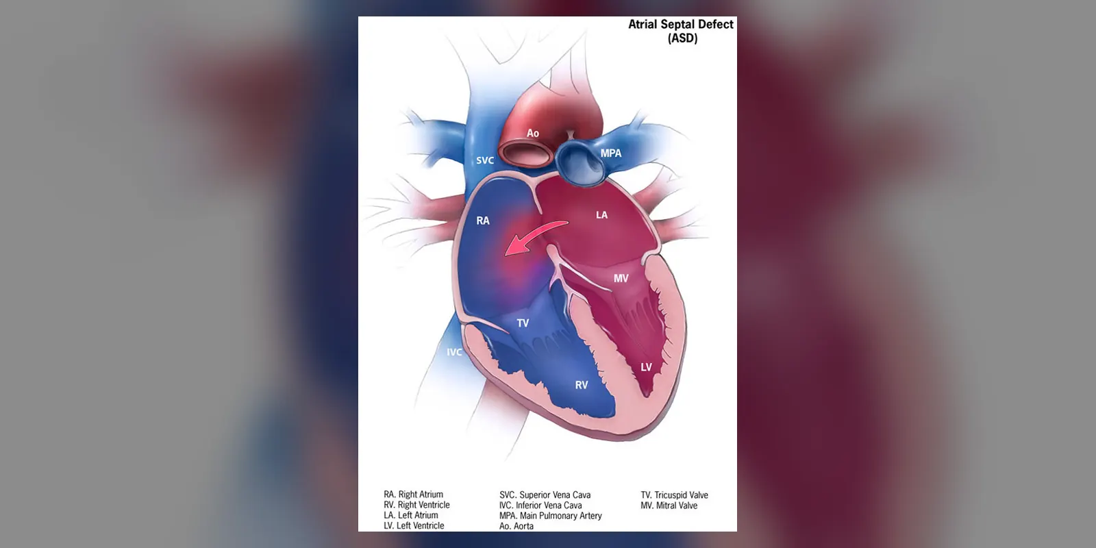
An atrial septal defect (ASD) is a hole in the wall (septum) that separates the two upper chambers of the heart, the left and right atria. It is present from birth and is one of the most common congenital heart defects. Because it often causes no symptoms in childhood, an ASD may not be discovered until adulthood.
Through the hole, oxygen-rich blood passes from the left atrium to the right, so the right side of the heart and the lungs handle extra blood. Over years, this extra load can enlarge the right side of the heart, cause abnormal heart rhythms and raise pressure in the lungs. Small ASDs may close on their own in childhood, while larger ones are usually closed with a catheter device or surgery.
When to get urgent help
Seek emergency care for stroke symptoms, severe breathlessness, chest pain or fainting.
Causes and risk factors
- Developmental: the atrial septum does not form fully before birth
- Genetic factors: more common in some families and in conditions such as Down syndrome and Holt-Oram syndrome
- Maternal factors during pregnancy: rubella, diabetes, smoking, alcohol and some medicines
How it develops (pathophysiology)
Normal heart development and circulation
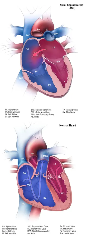
Before birth, blood bypasses the lungs, partly through an opening between the atria called the foramen ovale. After birth, as the lungs expand, pressure in the left atrium rises and a flap closes the foramen ovale. Normally the atrial septum is complete, so blood from the body (returning to the right atrium) and from the lungs (returning to the left atrium) stays separate.
What goes wrong
In an ASD, part of the septum fails to form:
- Secundum ASD (most common): a hole in the middle of the septum, where the foramen ovale is
- Primum ASD: low in the septum, often with mitral valve problems
- Sinus venosus ASD: near where the large veins enter the heart, often with abnormal lung vein drainage
- Coronary sinus defect: rare
Consequences
- Left-to-right shunt: pressure is normally slightly higher in the left atrium, and the right ventricle is more compliant, so blood flows from left to right through the hole.
- Volume overload: the right atrium, right ventricle and pulmonary arteries carry more blood than normal. Over years, they enlarge.
- Arrhythmias: stretching of the atria leads to atrial fibrillation and flutter, especially after age 40.
- Pulmonary hypertension: in a minority, long-term increased lung blood flow damages lung blood vessels, raising pressure.
- Eisenmenger syndrome: rarely, very high lung pressure reverses the shunt (right to left), sending oxygen-poor blood to the body and causing cyanosis. This is now rare with earlier detection.
- Paradoxical embolism: clots from the veins can cross the hole and cause a stroke.
Why the symptoms happen
- Breathlessness and tiredness on exertion result from the volume load and reduced efficiency.
- Palpitations reflect atrial arrhythmias.
- Frequent chest infections in children relate to increased lung blood flow.
- Heart murmur is caused by increased flow across the pulmonary valve, with a characteristic split second heart sound.
- Swollen legs may occur with right heart failure.
How treatment acts on the mechanism
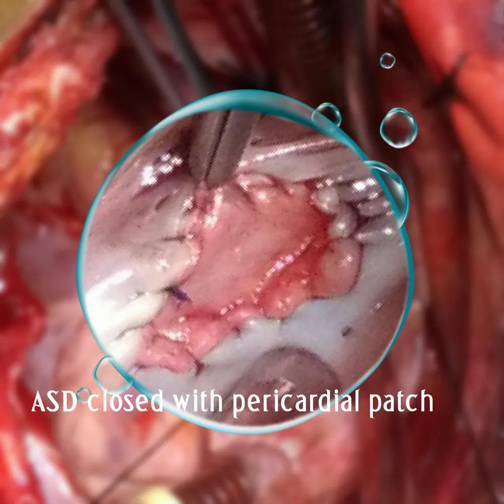
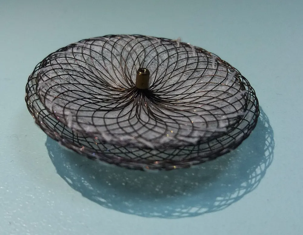
Closing the defect stops the shunt, allowing the right heart to shrink back towards normal size and reducing arrhythmia and pulmonary hypertension risk. Catheter device closure places an umbrella-like device through the hole via a vein. Surgical closure is used for defects unsuitable for devices, such as primum and sinus venosus ASDs.
Types
- Secundum ASD
- Primum ASD
- Sinus venosus ASD
- Coronary sinus ASD
- Patent foramen ovale (PFO): a related but distinct condition, present in about a quarter of adults
Signs and symptoms
- Often no symptoms, especially in children
- Breathlessness on exercise
- Tiredness
- Palpitations
- Frequent chest infections in childhood
- Swollen legs, feet or abdomen in later stages
- Stroke from paradoxical embolism (rare)
How it is diagnosed
- Heart murmur and fixed splitting of the second heart sound on examination
- ECG: may show right heart strain or arrhythmias
- Chest X-ray: may show an enlarged heart and prominent lung vessels
- Echocardiogram, including with bubble contrast
- Transoesophageal echocardiogram to measure the defect and plan closure
- Cardiac MRI or CT for sinus venosus defects
- Cardiac catheterisation to measure lung pressures in some cases
Possible complications
- Right heart enlargement and heart failure
- Atrial fibrillation and flutter
- Pulmonary hypertension
- Stroke from paradoxical embolism
- Eisenmenger syndrome (rare)
Goals of treatment
Treatment aims to close significant defects before complications develop, manage arrhythmias and pulmonary hypertension, and prevent stroke.
Medicines
- Diuretics for fluid overload
- Rate or rhythm control medicines for arrhythmias
- Anticoagulants for atrial fibrillation or after stroke
- Pulmonary hypertension medicines in selected cases
Other treatments
- Watchful waiting for small defects in children, many of which close on their own
- Catheter (transcatheter) device closure for suitable secundum ASDs
- Surgical closure for large, primum or sinus venosus defects
- Catheter ablation for arrhythmias
Living with it and prevention
ASDs cannot be prevented, but in pregnancy, avoiding alcohol and smoking, managing diabetes and having rubella immunity reduce the risk of congenital heart defects.
After diagnosis:
- Attend regular cardiology follow-up
- Report palpitations or breathlessness
- Plan pregnancy with a specialist
Monitoring and follow-up
After closure, echocardiograms check device position and heart size. Long-term follow-up monitors for arrhythmias and pulmonary hypertension.
Outlook
Small ASDs often close in childhood or cause no problems. Large ASDs closed early have an excellent outcome. Closure in adulthood still improves symptoms and heart size, though arrhythmia risk may persist.
Special considerations
- Pregnancy: usually well tolerated with an uncomplicated ASD, but significant pulmonary hypertension makes pregnancy high-risk
- Children with Down syndrome: screened for heart defects
- Adults over 40: higher risk of arrhythmias